4,068 posters, 219 videos, 101 topics, 69 sessions, 14,365 authors, 6,128 institutions
ePostersLive by SciGen Technologies S.A. All rights reserved.
Alzheimer's Association International Conference 2026
July 12-15, 2026 | London, United Kingdom, and Online

4138
Deep Learning-derived Brain Age From 40,000+ Mri Reveals Disease-specific Aging Trajectories
Poster Presenter
Authors
Jacques-Yves Campion, Rushil Srirambhatla, Carmen Andreescu, Thomas Desmidt, Helmet Karim
Affiliations
Department of Psychiatry, University of Pittsburgh, UMR 1253, iBraiN, Université de Tours, Inserm, Tours, France, Centre Hospitalier Régional Universitaire (CHRU) de Tours, Tours, France, Department of Applied Mathematics and Statistics, Johns Hopkins University, Department of Psychiatry, University of Pittsburgh, UMR 1253, iBraiN, Université de Tours, Inserm, Tours, France, Centre Hospitalier Régional Universitaire (CHRU) de Tours, Tours, France, Department of Psychiatry, University of Pittsburgh, Department of Bioengineering, University of Pittsburgh
Part of Topic
P1-09 - Deep Learning-Derived Brain Age from 40,000+ MRI Reveals Disease-Specific Aging
What if a single brainscan could tell you how fast you'reneurologically aging? We trained an AI on over 40,000 MRIs across 46 cohorts to estimate brain age — and discovered thatsome neurological and psychiatric diseasesage the brain by more than 10 years beyondwhat's expected.
Background
Brain age is the estimated biological age of an individual's brain predicted from structural MRI.
Methods
Population
We assembled a large population with over 40,841 T1-weighted MRI scans from more over than 46 independent cohorts and 27,190 individuals. A multi-label tag-based diagnostic framework allowed representation of comorbidity and disease. OASIS-3 and Cam-CAN datasets are held out for independent testing. 80% of the rest are used for training and the other 20% for validation.
Preprocessing
We applied the same pipeline to each scan startingwith a bias field correction of the fieldbias with ANTS then a rigid spatial registration to a standardized image space. Critically, we did not do spatial normalization – just rigid alignment. Pre-processed scans of IXI were used to train a histogramnormalization. We rescaled intensities to a range of 0-1, ignoring the bottom and top 0.5% values and applied the trained histogram normalization. Weapplied a head mask (excluding face). Optionally, an augmentation was applied (flip, blur, noise, motion, ghosting or spike).
Model Architecture
We adapted the Simple Fully Convolutional Network. The last layers are modified to do a regression insteadof classification. The batch size was fixed to 16 withan accumulation gradient of 9 and we used a halfprecision. Best option is learning rate is 0.04, dopoutis 0.4, initial channel of 32 and SGD optimizer.
Performances
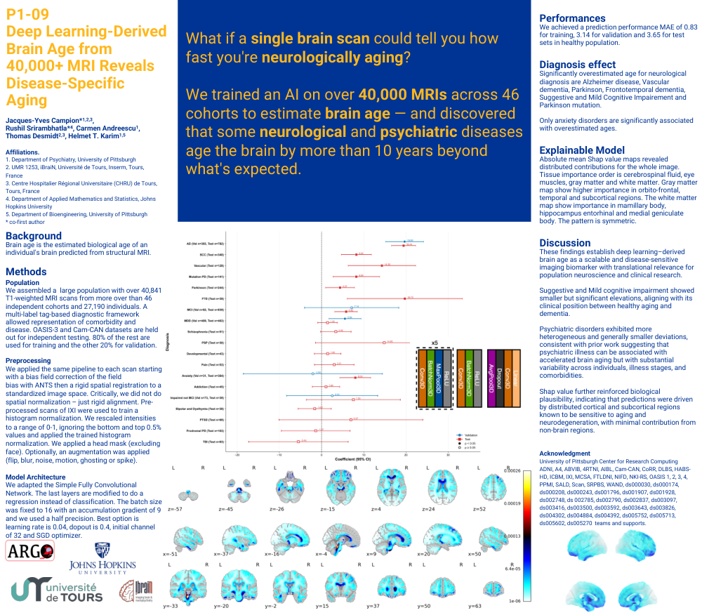
We achieved a prediction performance MAE of 0.83 for training, 3.14 for validation and 3.65 for test sets in healthy population.
Diagnosis effect
Significantly overestimated age for neurologicaldiagnosis are Alzheimer disease, Vascular dementia, Parkinson, Frontotemporal dementia, Suggestive and Mild Cognitive Impairement and Parkinson mutation.
Only anxiety disorders are significantly associatedwith overestimated ages.
Explainable Model
Absolute mean Shap value maps revealed distributedcontributions for the whole image. Tissue importance order is cerebrospinal fluid, eye muscles, gray matterand white matter. Gray matter map show higherimportance in orbito-frontal, temporal and subcortical regions. The white matter map show importance in mamillary body, hippocampus entorhinal and medialgeniculate body. The pattern is symmetric.
Discussion
These findings establish deep learning–derived brainage as a scalable and disease-sensitive imagingbiomarker with translational relevance for population neuroscience and clinical research.
Suggestive and Mild cognitive impairment showedsmaller but significant elevations, aligning with itsclinical position between healthy aging and dementia.
Psychiatric disorders exhibited more heterogeneousand generally smaller deviations, consistent with priorwork suggesting that psychiatric illness can beassociated with accelerated brain aging but withsubstantial variability across individuals, illnessstages, and comorbidities.
Shap value further reinforced biologicalplausibility, indicating that predictions were driven by distributed cortical and subcortical regions known to be sensitive to aging and neurodegeneration, withminimal contribution from non-brain regions.
Comments
Email is required but is not shown publicly.
Loading comments...