Loading...

4,068 posters, 219 videos, 101 topics, 69 sessions, 14,365 authors, 6,128 institutions

ePostersLive by SciGen Technologies S.A. All rights reserved.

Alzheimer's Association International Conference 2026

July 12-15, 2026 | London, United Kingdom, and Online

Alzheimer's Association International Conference 2026 banner

2253

Occipital Amyloid PET Deposition Across The AD Spectrum: A Multi-cohort Study
Poster Presenter

Alba Martos Sαnchez

Authors

Alba Martos Sαnchez, Wiesje Pelkmans, David Vállez García, Ana Harris Martínez, Joseph Therriault, Theresa M. Harrison, Gill Farrar, Juan Domingo Gispert, Frederik Barkhof, Sylvia Villeneuve, Juan Fortea, Pedro Rosa-Neto, Susan M. Landau, Lyduine E. Collij, Gemma Salvadó

Affiliations

1 Barcelonaβeta Brain Research Center (BBRC), Pasqual Maragall Foundation, Barcelona, Spain. 2 Universitat de Barcelona (UB), Barcelona, Spain. 3 Hospital del Mar Medical Research Institute (IMIM), Barcelona, Spain., 1 Barcelonaβeta Brain Research Center (BBRC), Pasqual Maragall Foundation, Barcelona, Spain. 3 Hospital del Mar Medical Research Institute (IMIM), Barcelona, Spain., 1 Barcelonaβeta Brain Research Center (BBRC), Pasqual Maragall Foundation, Barcelona, Spain. 3 Hospital del Mar Medical Research Institute (IMIM), Barcelona, Spain. 4 Centro de Investigación Biomédica en Red Bioingeniería, Biomateriales y Nanomedicina (CIBER‐BBN), Instituto de Salud Carlos III, Madrid, Spain, 1 Barcelonaβeta Brain Research Center (BBRC), Pasqual Maragall Foundation, Barcelona, Spain., 5 Translational Neuroimaging Laboratory, McGill University Research Centre for Studies in Aging, McConnell Brain Imaging Centre (BIC), Montréal Neurological Institute, Montréal, Québec, Canada. 6 Department of Neurology and Neurosurgery, McGill University, Montréal, Québec, Canada., 7 Neuroscience Department, University of California, Berkeley, CA, USA., 8 The Grove Centre, White Lion Road Buckinghamshire, GE HealthCare, Amersham, UK., 9 Queen Square Multiple Sclerosis Centre, Department of Neuroinflammation, UCL Queen Square Institute of Neurology, Faculty of Brain Sciences, University College London, London, UK; Centre for Medical Image Computing, University College London, London, UK. 10 Department of Radiology & Nuclear Medicine, Amsterdam UMC, Vrije Universiteit, Amsterdam, Netherlands., 11 Department of Psychiatry, McGill University, Montréal, QC, Canada. 12 Douglas Mental Health University Institute, Montreal, QC, Canada., 13 Sant Pau Memory Unit, Hospital de la Santa Creu i Sant Pau - Biomedical Research Institute Sant Pau, Barcelona, Spain. 14 Centro de Investigación Biomédica en Red de Enfermedades Neurodegenerativas, CIBERNED, Madrid, Spain; Barcelona Down Medical Center, Fundació Catalana Síndrome de Down, Barcelona, Spain., 12 Douglas Mental Health University Institute, Montreal, QC, Canada. 15 McGill University Research Centre for Studies in Aging, Montreal, QC, Canada; McGill University, Montreal, QC, Canada; Montreal Neurological Institute, Montreal, QC, Canada., 10 Department of Radiology & Nuclear Medicine, Amsterdam UMC, Vrije Universiteit, Amsterdam, Netherlands. 16 Amsterdam Neuroscience, Brain Imaging, Amsterdam, The Netherlands. 17 Clinical Memory Research Unit, Department of Clinical Sciences, Lund University, Malmö, Sweden., 1 Barcelonaβeta Brain Research Center (BBRC), Pasqual Maragall Foundation, Barcelona, Spain. 3 Hospital del Mar Medical Research Institute (IMIM), Barcelona, Spain. 17 Clinical Memory Research Unit, Department of Clinical Sciences, Lund University, Malmö, Sweden.

Part of Topic

Background: 

•In vivo amyloid-β (Aβ) PET assessment is typically done in the frontal, temporal, and parietal cortices1
•Emerging evidence shows Aβ can also accumulate in the Occipital lobe, a region currently excluded from standard quantification and visual reads2
•Investigating regions beyond typical accumulation sites may help explain, in part, the heterogeneity observed in AD through distinct disease subtypes2-6
•The extent to which Occipital Aβ deposition influences clinical progression and trial eligibility in preclinical stages remains poorly understood

Aim: 

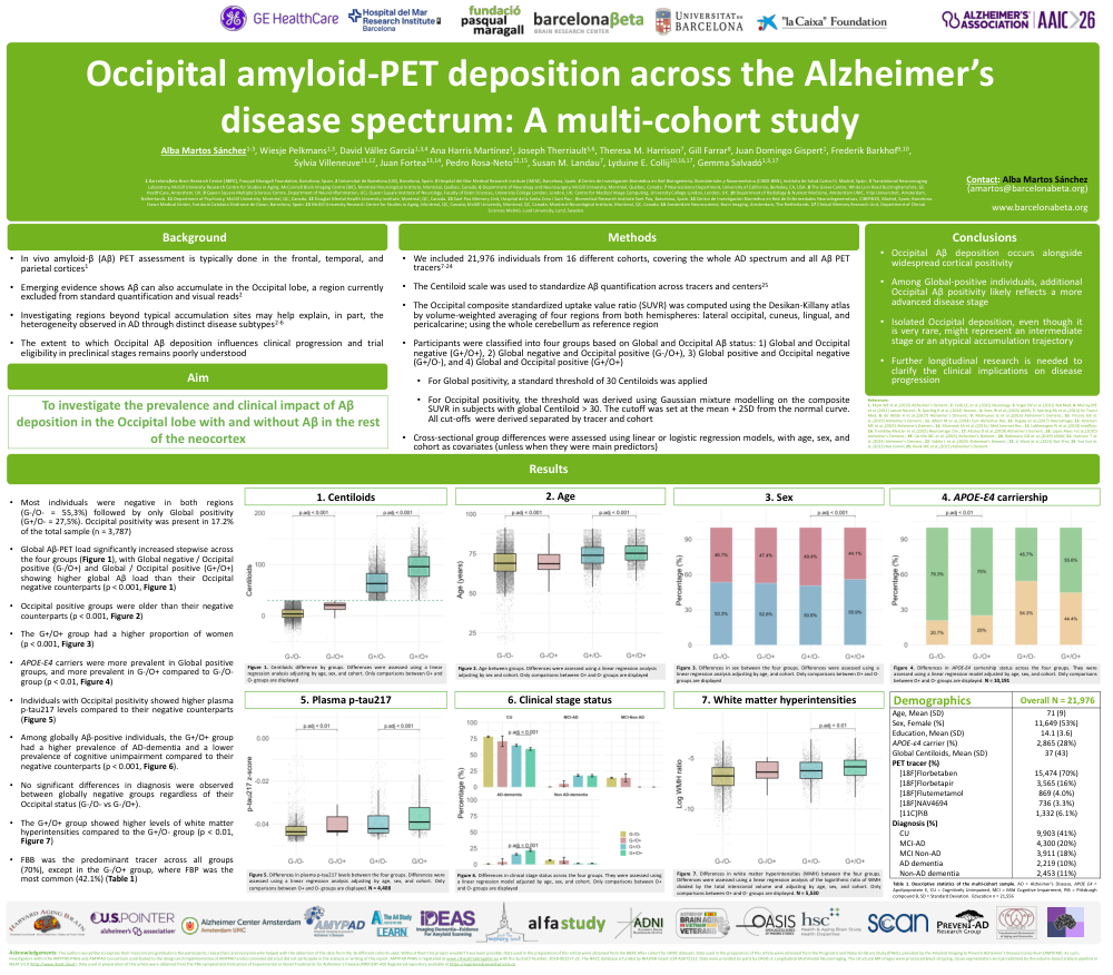
To investigate the prevalence and clinical impact of Aβ deposition in the Occipital lobe with and without Aβ in the rest of the neocortex

Methods: 

•We included 21,976 individuals from 16 different cohorts, covering the whole AD spectrum and all Aβ PET tracers7-24
•The Centiloid scale was used to standardize Aβ quantification across tracers and centers25
•The Occipital composite standardized uptake value ratio (SUVR) was computed using the Desikan-Killany atlas by volume-weighted averaging of four regions from both hemispheres: lateral occipital, cuneus, lingual, and pericalcarine; using the whole cerebellum as reference region
•Participants were classified into four groups based on Global and Occipital Aβ status: 1) Global and Occipital negative (G+/O+), 2) Global negative and Occipital positive (G-/O+), 3) Global positive and Occipital negative (G+/O-), and 4) Global and Occipital positive (G+/O+)
•For Global positivity, a standard threshold of 30 Centiloids was applied
•For Occipital positivity, the threshold was derived using Gaussian mixture modelling on the composite SUVR in subjects with global Centiloid > 30. The cutoff was set at the mean + 2SD from the normal curve. All cut-offs  were derived separated by tracer and cohort
•Cross-sectional group differences were assessed using linear or logistic regression models, with age, sex, and cohort as covariates (unless when they were main predictors)

Results: 
•Most individuals were negative in both regions (G-/O- = 55,3%) followed by only Global positivity (G+/O- = 27,5%). Occipital positivity was present in 17.2% of the total sample (n = 3,787)
•Global Aβ-PET load significantly increased stepwise across the four groups (Figure 1), with Global negative / Occipital positive (G-/O+) and Global / Occipital positive (G+/O+) showing higher global Aβ load than their Occipital negative counterparts (p < 0.001, Figure 1)
•Occipital positive groups were older than their negative counterparts (p < 0.001, Figure 2)
•The G+/O+ group had a higher proportion of women (p < 0.001, Figure 3)
•APOE-Ɛ4 carriers were more prevalent in Global positive groups, and more prevalent in G-/O+ compared to G-/O- group (p < 0.01, Figure 4)
•Individuals with Occipital positivity showed higher plasma p-tau217 levels compared to their negative counterparts (Figure 5)
•Among globally Aβ-positive individuals, the G+/O+ group had a higher prevalence of AD-dementia and a lower prevalence of cognitive unimpairment compared to their negative counterparts (p < 0.001, Figure 6).
•No significant differences in diagnosis were observed between globally negative groups regardless of their Occipital status (G-/O- vs G-/O+).
•The G+/O+ group showed higher levels of white matter hyperintensities compared to the G+/O- group (p < 0.01, Figure 7)
FBB was the predominant tracer across all groups (70%), except in the G-/O+ group, where FBP was the most common (42.1%) (Table 1)
 
Conclusion: 
•Occipital Aβ deposition occurs alongside widespread cortical positivity
•Among Global-positive individuals, additional Occipital Aβ positivity likely reflects a more advanced disease stage
•Isolated Occipital deposition, even though it is very rare, might represent an intermediate stage or an atypical accumulation trajectory
•Further longitudinal research is needed to clarify the clinical implications on disease progression

Comments

Email is required but is not shown publicly.

Loading comments...

aaic2026 banner
Copyright © 2015-2026, ePostersLive® SciGen is a registered trademark of SciGen Technologies S.A. Patent pending.

This website and third-party tools we use rely on cookies for the best user experience. By selecting "I agree", you agree to cookie usage as described in our Privacy Policy.