4,068 posters, 219 videos, 101 topics, 69 sessions, 14,365 authors, 6,128 institutions
ePostersLive by SciGen Technologies S.A. All rights reserved.
Alzheimer's Association International Conference 2026
July 12-15, 2026 | London, United Kingdom, and Online

10860
Multimodal Analysis Of Csf Biomarkers And Brain Morphometry In Scd/mci Stratified By Alzheimer’S Disease Pathophysiological Status
Poster Presenter
Authors
Part of Topic
Multimodal Analysis of CSF Biomarkers and Brain Morphometry in SCD/MCI Stratified by Alzheimer’s Disease Pathophysiological Status
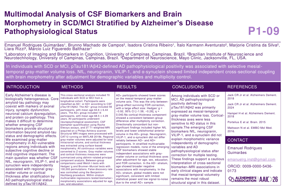
Introduction: Early Alzheimer’s disease is biologically heterogeneous. Core amyloid-tau pathology may coexist with markers of axonal injury, synaptic dysfunction, neuronal calcium dysregulation, and protein co-pathology. This makes it difficult to determine whether emerging CSF biomarkers provide structural information beyond amyloid-tau status and demographic effects. We examined gray-matter morphometry in AD-vulnerable regions among individuals with subjective cognitive decline or mild cognitive impairment. The main question was whether CSF NfL, neurogranin, VILIP-1, and α-synuclein were independently associated with regional gray-matter volume or cortical thickness after stratification by AD pathophysiological status defined by pTau181/Aβ42.
Methods: This cross-sectional analysis included 70 individuals with SCD or MCI from a longitudinal cohort. Participants were classified as AD− or AD+ according to CSF pTau181/Aβ42. The AD− group included 56 participants, with mean age 64.6 ± 6.44 years. The AD+ group included 14 participants, with mean age 68.5 ± 4.29 years. All participants underwent neuropsychological assessment, CSF biomarker quantification using Roche Elecsys assays, and 3T T1-weighted MRI acquired on a Philips Achieva scanner. Structural MRI images were processed with CAT12 running on MATLAB 2019b. Regional gray-matter volumes were adjusted for total intracranial volume, and cortical thickness was extracted using surface-based morphometry. All continuous variables were converted to Z-scores. Gray-matter volumes and cortical-thickness measures were summarized using oblimin-rotated principal component analysis. Between-group comparisons used Welch t-tests with Hedges’ g and Brunner–Munzel tests with 100,000 permutations. False discovery rate was controlled using the Benjamini–Hochberg procedure. Within-stratum multivariable regressions tested biomarker–morphometry associations adjusted for age, sex, and education.
Results: AD+ participants showed lower scores on the mesial-temporal gray-matter volume axis. This was the only between-group effect surviving FDR correction, with a large effect size: Hedges’ g = −0.92, 95% CI [−1.58, −0.28], q = 0.040.No cortical-thickness component showed a consistent between-group difference after multiplicity correction. Directionally concordant but non-significant findings included higher NfL levels and lower orbitofrontal-anterior volume in the AD+ group. Neurogranin, VILIP-1, and α-synuclein did not differ reliably between AD− and AD+ participants. In stratified multivariable regression models, none of the emerging CSF biomarkers showed stable independent associations with gray-matter volume or cortical-thickness axes after adjustment for age, sex, education, and FDR correction. In the AD− stratum, age showed broader negative associations with morphometry. In the AD+ stratum, global models were not significant, consistent with limited statistical power and low signal-to-noise due to the small AD+ sample.
Conclusion: Among individuals with SCD or MCI, AD pathophysiological positivity defined by pTau181/Aβ42 was primarily expressed as mesial-temporal gray-matter volume loss. Cortical-thickness axes were less sensitive to AD status in this sample.The emerging CSF biomarkers NfL, neurogranin, VILIP-1, and α-synuclein did not explain morphometric variance independently of demographic variables and AD pathophysiological status after correction for multiple testing. These findings support a cautious interpretation of cross-sectional biomarker–MRI associations in early clinical stages and indicate that mesial-temporal volumetry remains the most robust structural signal in this dataset.
Comments
Email is required but is not shown publicly.
Loading comments...