4,068 posters, 219 videos, 101 topics, 69 sessions, 14,365 authors, 6,128 institutions
ePostersLive by SciGen Technologies S.A. All rights reserved.
Alzheimer's Association International Conference 2026
July 12-15, 2026 | London, United Kingdom, and Online

10568
Sex Differences In Seeding And Propagation Of Amyloid, Tau And Neurodegeneration In AD Using A Bayesian Network Model
Poster Presenter
Authors
Vladimir Fonov, Seyyed Ali Hosseini, Lydia Trudel, Gleb Bezgin, Joseph Therriault, Arthur C. Macedo, Brandon J. Hall, Delphine Oliva-Lopez, Tevy Chan, Nesrine Rahmouni, Jenna Stevenson, Stijn Servaes, Serge Gauthier, Pedro Rosa-Neto, D. Louis Collins
Affiliations
McConnell Brain Imaging Centre, Montreal Neurological Institute, McGill University, Montreal, Canada, Translational Neuroimaging Laboratory, McGill University Research Centre for Studies in Aging, Douglas Hospital, McGill University, Montreal, Canada, McConnell Brain Imaging Centre, Montreal Neurological Institute, McGill University, Montreal, Canada, Translational Neuroimaging Laboratory, McGill University Research Centre for Studies in Aging, Douglas Hospital, McGill University, Montreal, Canada, Peter O'Donnell Jr Brain Institute, UT Southwestern Medical Centre, Dallas, TX, USA
Part of Topic
Background
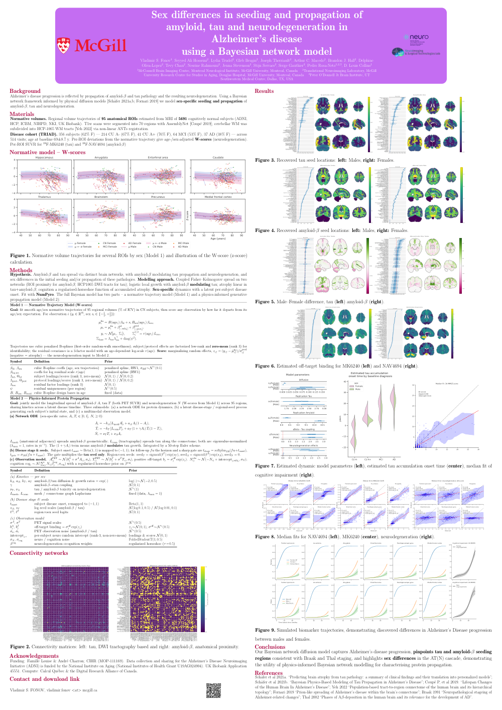
Alzheimer’s disease progression reflects the propagation of amyloid-β and tau pathology and resulting neurodegeneration. Using a Bayesian network framework informed by recent physical diffusion models [Schafer 2021a, 2021b, Fornari 2019], we model sex-specific seeding and propagation of amyloid, tau, and neurodegeneration.
Methods
We estimated trajectories of the normative regional volume change of 95 anatomical brain ROIs using MRI scans of 5691 healthy cognitively normal subjects without neurological abnormalities from ADNI, HCP, ICBM, NIHPD, NKI and UK Biobank. Each T1w MRI scan was segmented into 70 anatomical regions using AssemblyNet [Coupe], the ROI corresponding to cerebellar WM was subdevided regions corresponding to WM tracts from HCP-1065 fiber template [Yeh] using non-linear registration with ANTs.
Using data from TRIAD, (338 subjects, 481 visits, 242 CU , 62 MCI and 34 AD, mean age 69(8.5) years) we then calculated deviation from these expected volume trajectories of each ROI, in the form of an age- and sex-adjusted z-scored difference as a measure of neurodegeneration. In addition, for each ROI we calculated SUVR values for 18F-MK6240 (Tau) and 18F-NAV4694 (aBeta) PET tracers (Fig 1a), subtracted mean values from the cognitively normal young controls, and normalized all values between 0 and 1.
To model spread of Tau and aBeta protein we used The Fisher–Kolmogorov model explained in [Fornari 2019], where the local production rate alpha is modulated by the concentration of aBeta, local atrophy rate was modeled as a linear function of tau concentration, and the overall cognition was modelled as a linear function of regionalized accumulated atrophy with factorized random effect explaining inter-subject variability.
ROI proximity defined the connectivity matrix for aBeta and the HCP1064 DWI tract atlas was used to define Tau connectivity (Fig 1c).
We used the Bayesian modelling framework NumPyro to fit the model, and to estimate the initial seeding locations (and amounts) for Tau and aBeta as well as network spreading parameters.
Results
Results (Fig 2) show the recovered patterns of Tau seeding correspond to Braak [1991] stages I, II and III, while the amyloid seeding corresponds to stage B. Figure 3 shows how amyloid, tau and neurodegeneration occur in selected ROIs over time, differentially by sex.
Comments
Email is required but is not shown publicly.
Loading comments...