4,068 posters, 219 videos, 101 topics, 69 sessions, 14,365 authors, 6,128 institutions
ePostersLive by SciGen Technologies S.A. All rights reserved.
Alzheimer's Association International Conference 2026
July 12-15, 2026 | London, United Kingdom, and Online

9681
Plasma Aβ42/Aβ40 Identifies Preclinical Amyloid Changes Prior To PET Positivity
Poster Presenter
Authors
Noelia Fandos, María Pascual-Lucas, Leticia Sarasa, Jorge Loscos, Jose Terencio, Juan Pablo Tartari, Ángela Sanabria, Oscar Sotolongo-Grau, Amanda Cano, Lluís Tàrraga, Miren Gurruchaga, Agustín Ruíz, Xavier Montalban, Mercè Boada, Montserrat Alegret, Marta Marquié, José Antonio Allué
Affiliations
Grifols, Barcelona, Spain, Ace Alzheimer Center Barcelona -Universitat Internacional de Catalunya, Barcelona, Spain, Ace Alzheimer Center Barcelona -Universitat Internacional de Catalunya, Barcelona, Spain; Networking Research Center on Neurodegenerative Diseases (CIBERNED), Instituto de Salud Carlos III, Madrid, Spain, Ace Alzheimer Center Barcelona -Universitat Internacional de Catalunya, Barcelona, Spain; Networking Research Center on Neurodegenerative Diseases (CIBERNED), Instituto de Salud Carlos III, Madrid, Spain; Glenn Biggs Institute for Alzheimer's & Neurodegenerative Diseases, University of Texas Health Science Center, San Antonio, TX, USA,, Araclon Biotech - Grifols, Zaragoza, Spain,
Part of Topic
Background:
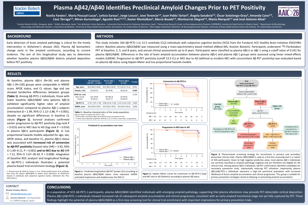
Early detection of brain amyloid pathology is critical for the timely intervention in Alzheimer’s disease (AD). Plasma Aβ biomarkers change early in the amyloid continuum, according to current evidence. The aim of this longitudinal study was to determine whether baseline plasma Aβ42/Aβ40 detects amyloid deposition before PET positivity.
Methods:
This study includes 164 Aβ-PET(−) (≤ 13.5 centiloids [CL]) individuals with subjective cognitive decline (SCD) from the Fundació ACE Healthy Brain Initiative (FACEHBI) cohort. Baseline plasma Aβ42/Aβ40 was measured using a mass-spectrometry based method (ABtest-MS, Araclon Biotech). Participants underwent 18F-Florbetaben PET at baseline, 2, 5, and 8 years, and annual clinical assessments up to 8 years. Participants were classified as plasma Aβ(+) or Aβ(−) using a cutoff value of 0.241 for plasma Aβ42/Aβ40. Differences in the rate of brain amyloid accumulation between plasma Aβ(+) and plasma Aβ(−) groups were assessed using linear mixed-effects models (LMEM). Progression to Aβ-PET positivity (cutoff 13.5 CL) or MCI due to AD (defined as incident MCI with concomitant Aβ-PET positivity) was evaluated based on plasma Aβ status using Kaplan-Meier and Cox proportional hazards models.
Results:
At baseline, plasma Aβ(+) (N=34) and plasma Aβ(−) (N=130) groups were comparable in MMSE score, APOE status, and CL values. Age and sex showed borderline differences between groups (Table 1). Among Aβ-PET(−) individuals, those with lower baseline Aβ42/Aβ40 ratio (plasma Aβ(+)) exhibited significantly higher rates of amyloid accumulation compared to plasma Aβ(−) subjects (interaction β = 1.99, 95% CI 1.12–2.86; P < 0.001), despite no significant differences in baseline CL values (Figure 1). Survival analyses confirmed earlier progression to Aβ-PET positivity (log-rank P = 0.015) and to MCI due to AD (log-rank P = 0.014) in plasma Aβ(+) participants (Figure 2). In Cox proportional hazards models adjusted for age, sex, APOE status, and baseline CL, plasma Aβ(+) status was associated with increased risk of conversion to Aβ-PET positivity (hazard ratio [HR] = 3.92, 95% CI 1.69–9.11, P = 0.001) and to MCI due to AD (HR = 7.11, 95% CI 1.67–30.29, P = 0.008). Integration of baseline ROC analysis1 and longitudinal findings in Aβ-PET(−) individuals illustrates a potential plasma-based trial enrichment strategy (Figure 3).
Conclusions:
In a population of SCD Aβ-PET(−) participants, plasma Aβ42/Aβ40 identified individuals with emerging amyloid pathology, supporting that plasma alterations may precede PET-detectable cortical deposition. Plasma Aβ(+)/Aβ-PET(−) individuals showed increased risk of subsequent amyloid accumulation and clinical progression, consistent with an early amyloid transitional stage not yet fully captured by PET. These findings highlight the potential of plasma Aβ42/Aβ40 as a first-step screening tool for clinical trial enrichment with important implications for primary prevention trials.
Comments
Email is required but is not shown publicly.
Loading comments...