4,068 posters, 219 videos, 101 topics, 69 sessions, 14,365 authors, 6,128 institutions
ePostersLive by SciGen Technologies S.A. All rights reserved.
Alzheimer's Association International Conference 2026
July 12-15, 2026 | London, United Kingdom, and Online

3277
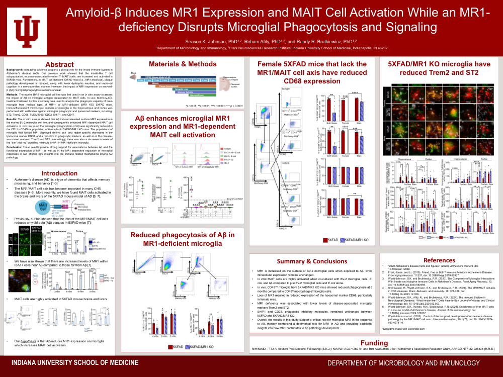
Amyloid Beta Induces MR1 Expression And MAIT Cell Activation While An MR1-deficiency Disrupts Microglial Phagocytosis And Signaling
Poster Presenter
Authors
Affiliations
Part of Topic
Video
Background: Increasing evidence supports a pivotal role for the innate immune system in Alzheimer's disease (AD). Our previous work showed that the innate-like T cell subpopulation, mucosal-associated invariant T (MAIT) cells, are increased and activated in 5XFAD mice. Furthermore, in MAIT cell-deficient 5XFAD mice (i.e., MR1-knockout), plaque pathology development is reduced, along with fewer dystrophic neurites, and improved cognition in a sex-dependent manner. However, the impact of MR1 expression on amyloid-β (Aβ) microglial phagocytosis remains unclear.
Methods: The murine BV-2 microglial cell line was first used in an in vitro assay to assess the impact of Aβ on microglial antigen presentation to MAIT cells. In vivo, Methoxy-X04 treatment followed by flow cytometry was used to analyze the phagocytic capacity of brain microglia from various ages of MR1+ or MR1-deficient (MR1 KO) 5XFAD mice. Immunofluorescent microscopic analysis of microglia in the hippocampus and cortex was performed with antibodies against microglial phagocytic and lysosomal markers, including: ST2, Trem2, CD68, TMEM106B, CD33, SHIP1, and CD47.
Results: The in vitro assays showed that Aβ induced elevated surface MR1 expression in the murine BV-2 microglial cell line, and consequently enhanced MR1-dependent MAIT cell activation. In vivo, we found that microglial phagocytosis of Aβ was significantly reduced in the CD11b+CD45low population of 6-month-old 5XFAD/MR1 KO mice. The populations of microglia that lacked MR1 displayed distinct sex- and region-specific decreases in the lysosomal marker CD68, and a reduction in phagocytic markers, as well as in the disease-associated markers, Trem2 and ST2. Interestingly, there was also a decrease in levels of the “don’t eat me” signaling molecule SHIP1 in MR1-deficient microglia.
Conclusion: These results provide strong support for associations between Aβ and the functional expression of MR1, as well as in the MR1-dependent regulation of microglial responses in AD, offering new insights into the immune-related mechanisms driving AD pathology.
Comments
Email is required but is not shown publicly.
Loading comments...