4,068 posters, 219 videos, 101 topics, 69 sessions, 14,365 authors, 6,128 institutions
ePostersLive by SciGen Technologies S.A. All rights reserved.
Alzheimer's Association International Conference 2026
July 12-15, 2026 | London, United Kingdom, and Online

5166
Spatially Resolved Cell-type-specific Molecular Drivers Of Anterior Temporal Pole Neuropathology In FTLD-TDP Type C
Poster Presenter
Part of Topic
Spatially Resolved Cell-Type-Specific Molecular Drivers of Anterior Temporal Pole Neuropathology in FTLD-TDP Type C
Brian Druciak1,2, Thomas Watson1,2, Alex J. Edwards1,2, Elena Barbieri3,4, Allegra S. Kawles4,5, Ivan Ayala4, Tamar Gefen4,5, Changiz Geula4,6, M. Marsel
Mesulam1,4, David Gate1,2 1Ken and Ruth Davee Department of Neurology, Northwestern University Feinberg School of Medicine, Chicago, IL, USA 2Abrams Research Center on Neurogenomics, Northwestern University Feinberg School of Medicine, Chicago, IL, USA 3Department of Physical Medicine and Rehabilitation, Feinberg School of Medicine, Chicago, IL, USA 4Mesulam Institute for Cognitive Neurology and Alzheimer’s Disease; Department of Cell and Developmental Biology, Feinberg School of Medicine, Northwestern University, Chicago, IL, USA 5Department of Psychiatry and Behavioral Sciences, Feinberg School of Medicine, Northwestern University, Chicago, IL, USA 6Department of Cell and Developmental Feinberg School of Medicine, Northwestern University, Chicago, IL, USA
Abstract
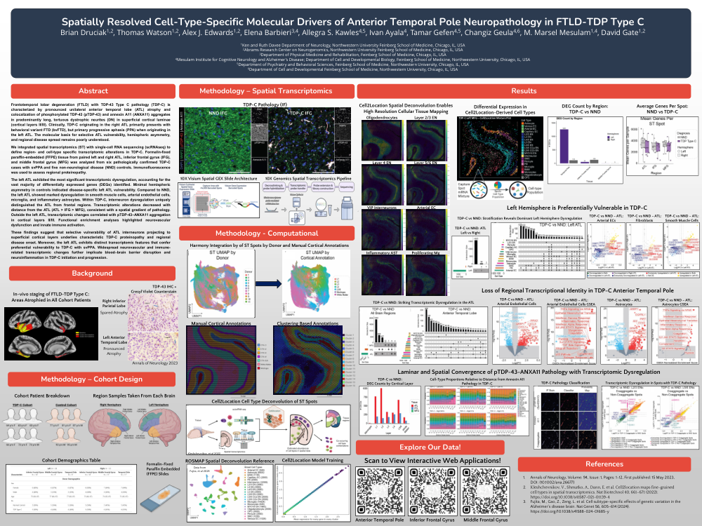
Frontotemporal lobar degeneration (FTLD) with TDP-43 Type C pathology (TDP-C) is characterized by pronounced unilateral anterior temporal lobe (ATL) atrophy and colocalization of phosphorylated TDP-43 (pTDP-43) and annexin A11 (ANXA11) aggregates in predominantly long, tortuous dystrophic neurites (DN) in superficial cortical laminae (cortical layers II/III). Clinically, TDP-C originating in the right ATL primarily presents with behavioral variant FTD (bvFTD), but primary progressive aphasia (PPA) when originating in the left ATL. The molecular basis for selective ATL vulnerability, hemispheric asymmetry, and regional disease spread remains poorly understood. We integrated spatial transcriptomics (ST) with single-cell RNA sequencing (scRNAseq) to define region- and cell-type specific transcriptomic alterations in TDP-C. Formalin-fixed paraffin-embedded (FFPE) tissue from paired left and right ATL, inferior frontal gyrus (IFG), and middle frontal gyrus (MFG) was analyzed from six pathologically confirmed TDP-C cases with svPPA and five non-neurological disease (NND) controls. Immunofluorescence was used to assess regional proteinopathy. The left ATL exhibited the most significant transcriptomic dysregulation, accounting for the vast majority of differentially expressed genes (DEGs) identified. Minimal hemispheric asymmetry in controls indicated disease-specific left ATL vulnerability. Compared to NND, the left ATL showed marked dysregulation in smooth muscle cells, arterial endothelial cells, microglia, and inflammatory astrocytes. Within TDP-C, interneuron dysregulation uniquely distinguished the ATL from frontal regions. Transcriptomic alterations decreased with distance from the ATL (ATL > IFG > MFG), consistent with a spatial gradient of pathology. Outside the left ATL, transcriptomic changes correlated with pTDP-43–ANXA11 aggregation in cortical layers II/III. Functional enrichment analyses highlighted neurovascular dysfunction and innate immune activation. These findings suggest that selective vulnerability of ATL interneurons projecting to superficial cortical layers underlies characteristic TDP-C proteinopathy and regional disease onset. Moreover, the left ATL exhibits distinct transcriptomic features that confer preferential vulnerability to TDP-C with svPPA. Widespread neurovascular and immune related transcriptomic changes further implicate blood–brain barrier disruption and neuroinflammation in TDP-C initiation and progression.
Comments
Email is required but is not shown publicly.
Loading comments...