4,068 posters, 219 videos, 101 topics, 69 sessions, 14,365 authors, 6,128 institutions
ePostersLive by SciGen Technologies S.A. All rights reserved.
Alzheimer's Association International Conference 2026
July 12-15, 2026 | London, United Kingdom, and Online

10746
Predicting Hippocampal Atrophy With PET And CSF Biomarkers In Alzheimer’S Disease: A Cross-sectional Study
Poster Presenter
Authors
Bernardo Cimirro, Fernando Lazzaretti, Fabricio Garrafiel, Cristiano Aguzzoli, Lucas Schilling
Affiliations
School of Medicine, Pontifícia Universidade Católica do Rio Grande do Sul, Porto Alegre, Brazil., Brain Institute of the Pontifical Catholic University of Rio Grande do Sul, Porto Alegre, Brazil.
Part of Topic
Background: Alzheimer’s disease is characterized by multiple pathological processes involving amyloid-β (Aβ) and tau. It remains uncertain which marker most reliably reflects the degree of hippocampal atrophy. We aimed to identify which biomarker best predicts hippocampal atrophy.
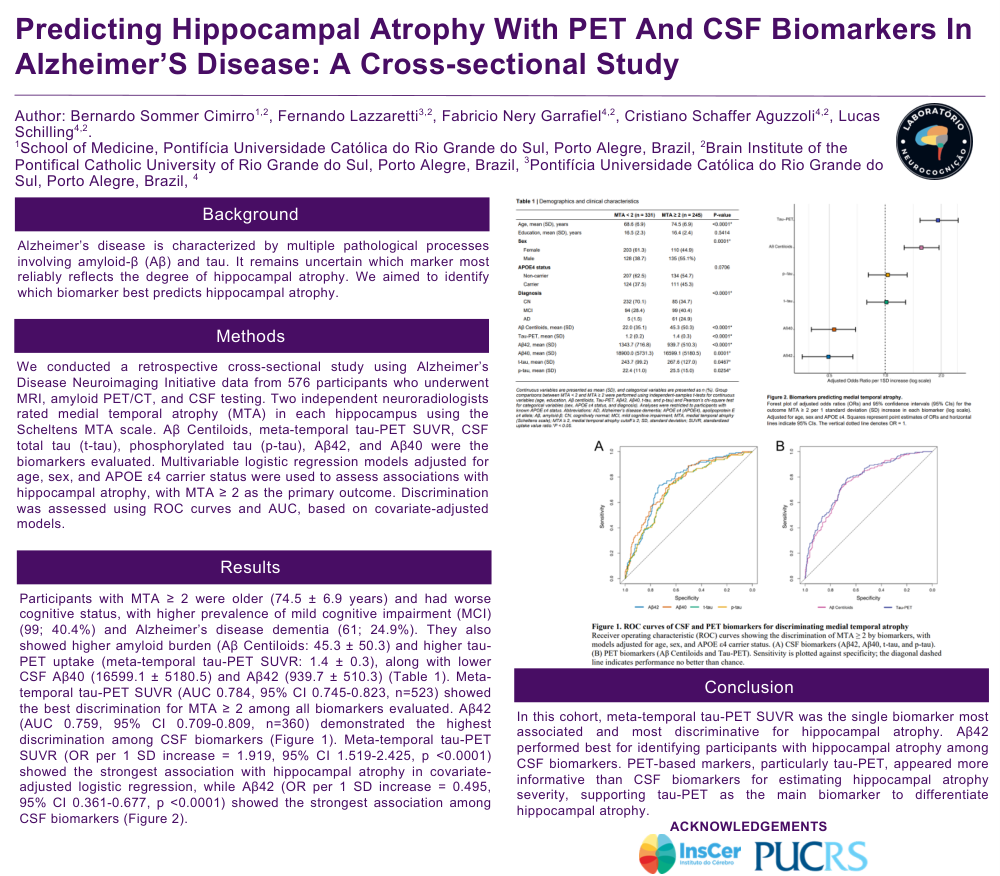
Methods: We conducted a retrospective cross-sectional study using Alzheimer’s Disease Neuroimaging Initiative data from 576 participants who underwent MRI, amyloid PET/CT, and CSF testing. Two independent neuroradiologists rated medial temporal atrophy (MTA) in each hippocampus using the Scheltens MTA scale. Aβ Centiloids, meta-temporal tau-PET SUVR, CSF total tau (t-tau), phosphorylated tau (p-tau), Aβ42, and Aβ40 were the biomarkers evaluated. Multivariable logistic regression models adjusted for age, sex, and APOE ε4 carrier status were used to assess associations with hippocampal atrophy, with MTA ≥ 2 as the primary outcome. Discrimination was assessed using ROC curves and AUC, based on covariate-adjusted models.
Results: Participants with MTA ≥ 2 were older (74.5 ± 6.9 years) and had worse cognitive status, with higher prevalence of mild cognitive impairment (MCI) (99; 40.4%) and Alzheimer’s disease dementia (61; 24.9%). They also showed higher amyloid burden (Aβ Centiloids: 45.3 ± 50.3) and higher tau-PET uptake (meta-temporal tau-PET SUVR: 1.4 ± 0.3), along with lower CSF Aβ40 (16599.1 ± 5180.5) and Aβ42 (939.7 ± 510.3) (Table 1). Meta-temporal tau-PET SUVR (AUC 0.784, 95% CI 0.745-0.823, n=523) showed the best discrimination for MTA ≥ 2 among all biomarkers evaluated. Aβ42 (AUC 0.759, 95% CI 0.709-0.809, n=360) demonstrated the highest discrimination among CSF biomarkers (Figure 1). Meta-temporal tau-PET SUVR (OR per 1 SD increase = 1.919, 95% CI 1.519-2.425, p <0.0001) showed the strongest association with hippocampal atrophy in covariate-adjusted logistic regression, while Aβ42 (OR per 1 SD increase = 0.495, 95% CI 0.361-0.677, p <0.0001) showed the strongest association among CSF biomarkers (Figure 2).
Conclusion: In this cohort, meta-temporal tau-PET SUVR was the single biomarker most associated and most discriminative for hippocampal atrophy. Aβ42 performed best for identifying participants with hippocampal atrophy among CSF biomarkers. PET-based markers, particularly tau-PET, appeared more informative than CSF biomarkers for estimating hippocampal atrophy severity, supporting tau-PET as the main biomarker to differentiate hippocampal atrophy.
Comments
Email is required but is not shown publicly.
Loading comments...