4,068 posters, 219 videos, 101 topics, 69 sessions, 14,365 authors, 6,128 institutions
ePostersLive by SciGen Technologies S.A. All rights reserved.
Alzheimer's Association International Conference 2026
July 12-15, 2026 | London, United Kingdom, and Online

496
Hippocampal CA3 Hyperexcitability Is Associated With Increased Sodium Channel Nav1.3 In A Mouse Model Of Early Alzheimer's Disease
Part of Topic
Video
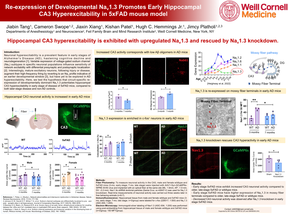
Re-expression of Developmental Nav1.3 Promotes Early Hippocampal CA3 Hyperexcitability in 5xFAD mouse model.
Hippocampal CA3 hyperexcitability is exhibited with upregulated Nav1.3 and rescued by Nav1.3 knockdown.
Hippocampal CA3 neuronal activity is increased in early AD mice.
Increased CA3 activity corresponds with low Aβ oligomers in AD mice.
Nav1.3 expression is enriched in c-fos+ neurons in early AD mice.
Nav1.3 is re-expressed on mossy fiber terminals in early AD mice.
Nav1.3 knockdown rescues CA3 hyperactivity in early AD mice.
Introduction: Neuronal hyperexcitability is a prevalent feature in early stages of Alzheimer's Disease (AD), hastening cognitive decline and neurodegeneration [1]. Variable expression of voltage-gated sodium channel (Nav) subtypes in specific neuronal populations influence sensitivity of network excitability with differential presynaptic and postsynaptic localization [2]. Interestingly, mature excitatory neurons, following injury or disease, augment their high-frequency firing by reverting to an Nav profile indicative of an earlier developmental window [3], but have yet to be explored in AD hyperexcitability. Here, we test the hypothesis that circuit-specific re-expression of developmentally dominant Nav1.3 potentiates hippocampal CA3 hyperexcitability in early stage of disease of 5xFAD mice, compared to both later-stage disease and non-AD controls.
Methods: Fiber Photometry: To measure neuronal activity in the CA3, male and female wildtype and 5xFAD mice (3-mo, early stage; 7-mo, late stage) were injected with AAV1-Syn-GCaMP6s-WPRE-SV40 virus and implanted with an optical fiber at the same site (ML: 1.9mm, AP: -1.9 mm, DV: -2.2 mm). Nav1.3α shRNA lentiviral particles (Santa Cruz, sc-42647-V) was used for Nav1.3 knockdown. Real-time measurements of neuronal activity was carried out three weeks later in free behaving animals (n=6-7). Immunofluorescence: Hippocampal tissue from male and female wildtype and 5xFAD mice (3-mo, early stage; 7-mo, late stage; n=3/group) were labeled for c-fos (226017, 1:500) and Nav1.3 (ASC-004, 1:200). Electron Microscopy: Immunogold-silver labeling of Nav1.3 (ASC-004, 1:200) was performed using post-embedding from hippocampal tissue of male and female wildtype and 5xFAD mice (n=3/group; 150 MFT/group).
Results: - Early stage 5xFAD mice exhibit increased CA3 neuronal activity compared to older, late-stage 5xFAD or wildtype mice. - Early stage 5xFAD mice have higher expression of Nav1.3 in mossy fiber terminals compared to older, late-stage 5xFAD or wildtype mice. - Decreased CA3 neuronal activity was observed after Nav1.3 knockdown in early stage 5xFAD mice.
Comments
Email is required but is not shown publicly.
Loading comments...