4,068 posters, 219 videos, 101 topics, 69 sessions, 14,365 authors, 6,128 institutions
ePostersLive by SciGen Technologies S.A. All rights reserved.
Alzheimer's Association International Conference 2026
July 12-15, 2026 | London, United Kingdom, and Online

9883
The Role Of Microglial Extracellular Vesicles In C1q-mediated Synaptic Pruning: Insights From Extracellular Vesicles From Brain Tissue
Poster Presenter
Authors
Martina Gabrielli, Giulia D'Arrigo, Giulia Cutugno, Maria Teresa Golia, Francesca Sironi, Marta Lombardi, Roberto Frigerio, Marina Cretich, Elisabetta Battocchio, Sara Francesca Colombo, Cristiana Barone, Emanuele Azzoni, Roberta Ghidoni, Caterina Bendotti, Rosa Chiara Paolicelli, Claudia Verderio
Affiliations
CNR Institute of Neuroscience, Vedano al Lambro, Italy; Current address: Nottingham Trent University, Nottingham, United Kingdom, CNR Institute of Neuroscience, Vedano al Lambro, Italy, Istituto di Ricerche Farmacologiche Mario Negri IRCCS, Milano, Italy, CNR Institute of Chemical Science and Technologies, Milano, Italy, University of Milano-Bicocca, Milano, Italy, IRCCS Istituto Centro San Giovanni di Dio Fatebenefratelli, Brescia, Italy, University of Lausanne, Lausanne, Switzerland
Part of Topic
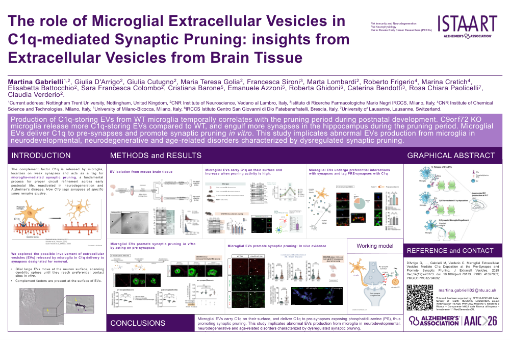
Background: The complement factor C1q is released by microglia, localizes on weak synapses and acts as a tag for microglia-mediated synaptic pruning, a fundamental process for proper circuit refinement across early postnatal life, reactivated in neurodegeneration and Alzheimer’s disease. However, how C1q tags synapses at specific times remains elusive. We explored the possible involvement of extracellular vesicles (EVs) released by microglia in C1q delivery to synapses designated for removal.
Methods: We optimized a protocol to extract large and small EVs from the interstitial fluid of mouse brain tissue. By Western Blot, TRPS, single molecules arrays (Simoa) and confocal imaging, we measured the amount, cell source and complement cargo of EVs extracted from WT postnatal pups and adult mice brains, and from C9orf72KO adult brains displaying enhanced microglia-mediated pruning. Then, we performed in vitro and ex vivo studies to inquire microglial EVs involvement in C1q deposition to the synapse and synaptic engulfment, using approaches to increase or reduce microglial EVs production by EVs supplementation C9orf72 silencing or pharmacological inhibition (GW4869).
Results: We show that microglia release more EVs carrying C1q in C9orf72KO adult mice, providing a link between microglial EVs production and synaptic removal. Moreover, we report that production of C1q carrying microglial EVs peaks during the pruning period in the early postnatal hippocampus (postnatal day P17), confirming a positive correlation between EVs production and synaptic removal in a physiological setting. In neuron-microglia co-cultures, microglial EVs, labelled by the fluorescent dye mCLING, make preferential contacts with synapses, deliver C1q to pre-synapses that externalize phosphatidylserine and promote synaptic removal. Interestingly, C9orf72 KO microglia engulf more synaptic terminals and decreases synaptic density to a greater extent compared to WT microglia whereas inhibition of EVs release by GW4869 restores normal pre-synaptic density, providing mechanistic evidence linking EVs release to synaptic remodeling.
Conclusion: This study identifies microglial EVs as delivery vehicles for C1q to synapses targeted for removal and implicates abnormal EVs production from microglia in neurodegeneration and age-related disorders characterized by dysregulated synaptic pruning. Analysis of specific subpopulations of brain EVs opens new perspectives to the understanding of EVs roles in physiology and pathology.
Comments
Email is required but is not shown publicly.
Loading comments...