4,068 posters, 219 videos, 101 topics, 69 sessions, 14,365 authors, 6,128 institutions
ePostersLive by SciGen Technologies S.A. All rights reserved.
Alzheimer's Association International Conference 2026
July 12-15, 2026 | London, United Kingdom, and Online

9319
Delineating White Matter In The Medial Temporal Lobe Using High-Resolution Diffusion MRI
Poster Presenter
Part of Topic
Introduction:
The temporal part of the cingulum bundle (CBT) and the fornix (FX) are two important white matter (WM) tracts within the medial temporal lobe (MTL) that are closely associated with cognitive functions, particularly memory.
Accurate delineation of these WM structures and characterization of their microstructural properties are essential for identifying and understanding neurodegeneration associated with normal aging and pathological processes. Due to their fine scale, reliable visualization of these tracts requires high-resolution diffusion MRI (DMRI) with moderate to high b-values 1.
This study aims to use an SNR-efficient, high-resolution DMRI acquisition technique, gSLIDER 2,3, to achieve 1 mm isotropic resolution and demonstrate its utility in delineating the CBT and FX.
Methods:
Data acquisition: Nine cognitively normal older subjects (4F/5M, 62~71 y/o) were scanned on a 3T GE scanner. Two DMRI scans were conducted, (1) a single-shell (b=1 ms/μm2) high-resolution DMRI protocol (1.0 mm isotropic) and (2) a two-shell (b=1 and 2 ms/μm2) moderate-resolution DMRI protocol (1.5 mm isotropic). Detailed parameters are listed in Table 1. Each subject was scanned twice (scans 1 and 2) to evaluate the scan-rescan reproducibility of the delineation of each WM tract.
Processing & analysis:
(1) The preprocessing of diffusion-weighted images (DWIs) includes denoising 4, Gibbs ringing artifacts 5, distortion correction 6, and motion correction 7.
(2) All preprocessed DWIs were registered to a T1-weighted (T1w) MPRAGE image, and probabilistic tractography was performed using FSL probtrackx2_gpu 8. The normalized fiber probability maps of CBT and FX were binarized by a percentage threshold of the normalized fiber probability of 1.5%, as well as fractional anisotropy (FA)≥0.2 and mean diffusivity (MD)≤1.5 μm2/ms (to mitigate partial volume effects from CSF and gray matter). The generated binary CBT and FX masks were used to estimate the the total voxel volume (Vol).
(3) The scan-rescan reproducibility was quantified using the Dice Similarity Coefficient (DSC), defined as the ratio between the overlapped volume and the average volume of the two scans: DSC=2×(Volscan1∩Volscan2)/(Volscan1+Volscan2). A DSC≥0.70 indicates good reproducibility, and 0.50≤DSC<0.70 indicates moderate reproducibility.
Results:
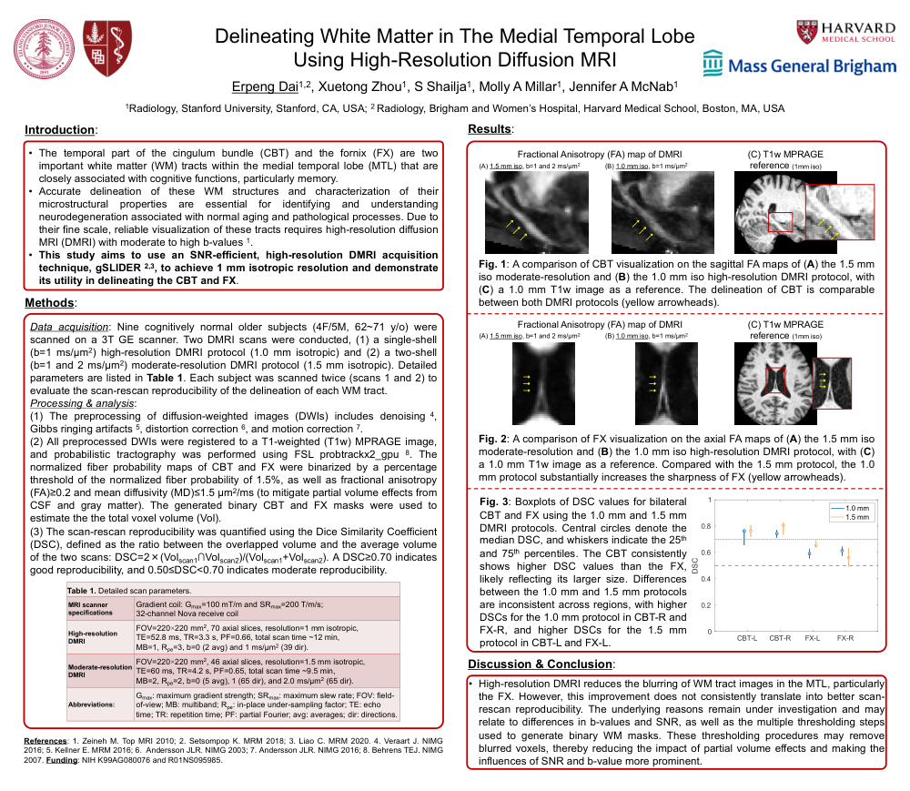
Fig. 1: A comparison of CBT visualization on the sagittal FA maps of (A) the 1.5 mm iso moderate-resolution and (B) the 1.0 mm iso high-resolution DMRI protocol, with (C) a 1.0 mm T1w image as a reference. The delineation of CBT is comparable between both DMRI protocols (yellow arrowheads).
Fig. 2: A comparison of FX visualization on the axial FA maps of (A) the 1.5 mm iso moderate-resolution and (B) the 1.0 mm iso high-resolution DMRI protocol, with (C) a 1.0 mm T1w image as a reference. Compared with the 1.5 mm protocol, the 1.0 mm protocol substantially increases the sharpness of FX (yellow arrowheads).
Fig. 3: Boxplots of DSC values for bilateral CBT and FX using the 1.0 mm and 1.5 mm DMRI protocols. Central circles denote the median DSC, and whiskers indicate the 25th and 75th percentiles. The CBT consistently shows higher DSC values than the FX, likely reflecting its larger size. Differences between the 1.0 mm and 1.5 mm protocols are inconsistent across regions, with higher DSCs for the 1.0 mm protocol in CBT-R and FX-R, and higher DSCs for the 1.5 mm protocol in CBT-L and FX-L.
Discussion & Conclusion:
High-resolution DMRI reduces the blurring of WM tract images in the MTL, particularly the FX. However, this improvement does not consistently translate into better scan-rescan reproducibility. The underlying reasons remain under investigation and may relate to differences in b-values and SNR, as well as the multiple thresholding steps used to generate binary WM masks. These thresholding procedures may remove blurred voxels, thereby reducing the impact of partial volume effects and making the influences of SNR and b-value more prominent.
References: 1. Zeineh M. Top MRI 2010; 2. Setsompop K. MRM 2018; 3. Liao C. MRM 2020. 4. Veraart J. NIMG 2016; 5. Kellner E. MRM 2016; 6. Andersson JLR. NIMG 2003; 7. Andersson JLR. NIMG 2016; 8. Behrens TEJ. NIMG 2007. Funding: NIH K99AG080076 and R01NS095985.
Comments
Email is required but is not shown publicly.
Loading comments...