516 posters, 59 topics, 63 sessions, 1,127 authors, 352 institutions
ePostersLive by SciGen Technologies S.A. All rights reserved.
2026 SOAP Annual Meeting
April 29 - May 3, 2026 | Montreal, Quebec Canada

2320759
Use of lumbar ultrasound in neuraxial anesthesia for a pregnant patient with Klippel–Trenaunay syndrome: a case report
Poster Presenter
Authors
Affiliations
Part of Topic
NEUROVASCULAR CHALLENGES
Lumbar ultrasound for neuraxial anesthesia in a parturient with Klippel–Trenaunay syndrome
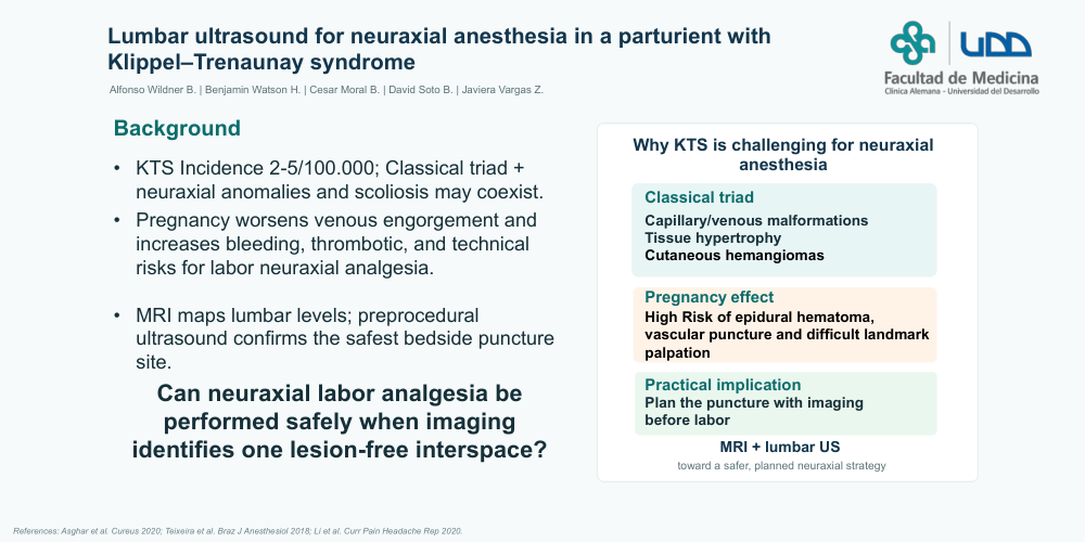
Klippel–Trenaunay syndrome (KTS) is a rare congenital disorder characterized by cutaneous hemangiomas, venous malformations, and tissue hypertrophy. It is often associated with neuraxial anomalies and scoliosis, complicating anesthesia. Pregnancy exacerbates these malformations, increasing bleeding and thrombotic risks. MRI in conjunction with lumbar ultrasound has emerged as a strategy that aids in the identification of anatomical structures, improves success rates, enhances patient satisfaction, and reduces the risk of complications. There are no previous Chilean reports on this association, and the combined use of MRI and lumbar ultrasound in obstetric patients with KTS has been sparingly documented internationally
We present a 40-year-old pregnant patient (37+5 weeks) with KTS and mild scoliosis. Her history included a prior complicated delivery involving intravascular epidural catheter placement. Given this history, lumbar MRI was performed, revealing anterior epidural space occupancy from L4 to S1 and venous prominence, while identifying the L2–L3 space as lesion-free. A pre-procedural lumbar ultrasound confirmed the patency of L2–L3 and provided depth measurements. Combined spinal-epidural (CSE) anesthesia was successfully performed at this level without vascular or neurological complications. Epidural PCEA was programmed achieving satisfactory analgesia.
Neuraxial ultrasound has established itself as a valuable tool for increasing the safety and efficacy of anesthesia in patients with complex anatomy or high vascular risk, such as those with KTS. This syndrome can be associated with vascular malformations within the neuraxis, increasing the risk of complications. In this context, planning based on imaging studies is essential.
MRI allows for the mapping of lesions and the selection of segments free of involvement, while lumbar ultrasound complements this information at the point of care, facilitating the identification of anatomical structures, depth estimation, and needle trajectory planning. The literature demonstrates that ultrasound improves vertebral level localization, increases the first-attempt success rate, and reduces failed attempts; this is particularly relevant in obstetrics, where the anatomical changes of pregnancy make the palpation of bony landmarks difficult
In this case, the combination of MRI and lumbar US allowed for the selection of a safe interspace and the optimization of the technique, minimizing risks and complications. Although it is not a substitute for MRI in ruling out intradural malformations, ultrasound provides practical advantages and additional safety at the time of puncture. Its systematic application in patients with KTS or other neuraxial vascular malformations could improve clinical outcomes, and this case contributes evidence to a field that is sparingly documented, especially at the national level