816 posters, 24 topics, 3,314 authors, 1,012 institutions
ePostersLive by SciGen Technologies S.A. All rights reserved.
SAGES 2026 Annual Meeting Emerging Technology Session
March 25-28, 2026 | Tampa, FL, USA

P663
IT TAKES A VILLAGE: MULTIDISCIPLINARY APPROACH TO RESTORATION OF LUMINAL CONTINUITY AFTER COMPLETE ESOPHAGOGASTRIC ANASTOMOTIC DEHISCENCE
Poster Presenter
Authors
Kaylie Nichols, Jussuf Kaifi, Charles Kitley, Joshua Landreneau
Affiliations
University of Missouri-Columbia School of Medicine, University of Missouri-Columbia Department of Cardiothoracic Surgery, University of Missouri-Columbia Department of Interventional Radiology, University of Missouri-Columbia Department of General Surgery
Part of Topic
Image Guided Interventions (Including Combined Radiologic / Endoscopic / Surgical Procedures)
IT TAKES A VILLAGE: MULTIDISCIPLINARY APPROACH TO RESTORATION OF LUMINAL CONTINUITY AFTER COMPLETE ESOPHAGOGASTRIC ANASTOMOTIC DEHISCENCE
Kaylie Nichols, BS, Jussuf Kaifi, MD, Charles Kitley, MD, Joshua Landreneau, MD
Department of Surgery, University of Missouri School of Medicine, Columbia, Missouri
INTRODUCTION
Esophageal anastomotic leak following esophagectomy carries significant morbidity and mortality. Complete anastomotic dehiscence poses a particular challenge, as conventional interventions such as stenting or endoluminal vacuum therapy are often infeasible. We present a case of complete esophagogastric anastomotic dehiscence successfully managed through multidisciplinary rendezvous recanalization.
CASE PRESENTATION
A 75-year-old male with gastroesophageal junction adenocarcinoma underwent an open transhiatal esophagectomy via upper midline laparotomy with left cervical esophagogastric anastomosis. His early postoperative course was complicated by cervical anastomotic leak and complete esophagogastric anastomotic dehiscence leading to formation of a cervical esophagocutaneous fistula.
PROCEDURES
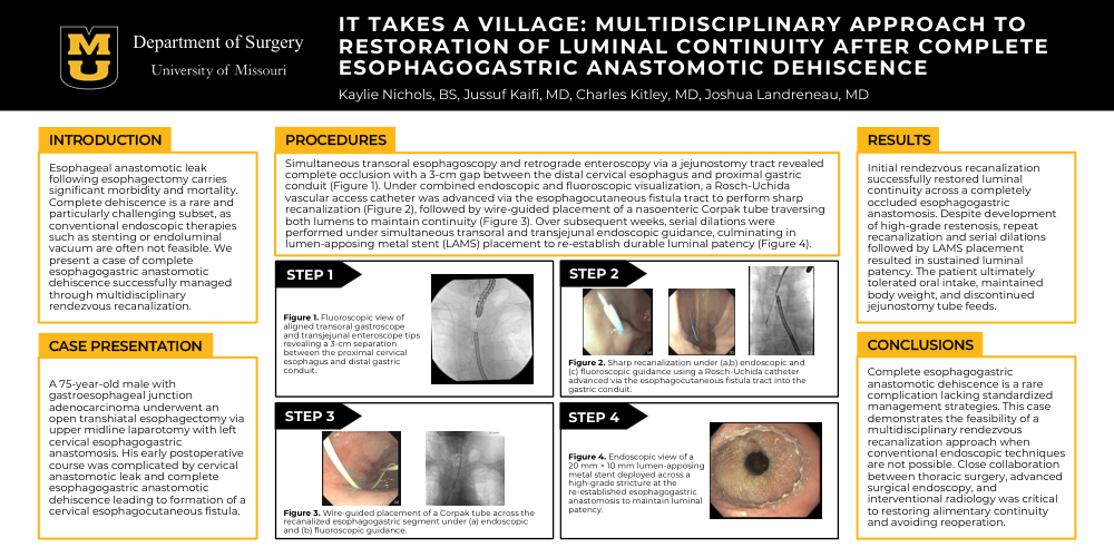
Simultaneous transoral esophagoscopy and retrograde enteroscopy via a jejunostomy tract revealed complete occlusion with a 3-cm gap between the distal cervical esophagus and proximal gastric conduit (Figure 1). Under combined endoscopic and fluoroscopic visualization, a Rosch-Uchida vascular access catheter was advanced via the esophagocutaneous fistula tract to perform sharp recanalization (Figure 2), followed by wire-guided nasoenteric Corpak tube placement traversing both lumens to maintain continuity (Figure 3). Over subsequent weeks, serial dilations were performed under simultaneous transoral and transjejunal endoscopic guidance, culminating in lumen-apposing metal stent (LAMS) placement to re-establish durable luminal patency (Figure 4).
RESULTS
Initial rendezvous recanalization successfully restored luminal continuity across a completely occluded esophagogastric anastomosis. Despite development of high-grade restenosis, repeat recanalization and serial dilations followed by LAMS placement resulted in sustained luminal patency. The patient ultimately tolerated oral intake, maintained body weight, and discontinued jejunostomy tube feeds.
CONCLUSIONS
Complete esophagogastric anastomotic dehiscence is a rare complication lacking standardized management strategies. This case demonstrates the feasibility of a multidisciplinary rendezvous recanalization approach when conventional endoscopic techniques are not possible. Close collaboration between thoracic surgery, advanced surgical endoscopy, and interventional radiology was critical to restoring alimentary continuity and avoiding reoperation.