816 posters, 24 topics, 3,314 authors, 1,012 institutions
ePostersLive by SciGen Technologies S.A. All rights reserved.
SAGES 2026 Annual Meeting Emerging Technology Session
March 25-28, 2026 | Tampa, FL, USA

P608
Hypoperfusion From Necrotizing Soft Tissue Infection Causing Esophageal Necrosis
Poster Presenter
Part of Topic
Foregut
HYPOPERFUSION FROM NECROTIZING SOFT TISSUE INFECTION CAUSING ESOPHAGEAL NECROSISHilla R Katz-Lichtenstein, MD; Rachel Stern, MD; Jonathan Bakhshi; Kirill Antonov, MD; Tara Ranjbar, MD; Tamar Gordis, MD; Manuel Villa Sanchez, MD; Indraneil Mukherjee, MBBS, MD; Northwell Health (SSO)
Introduction: Esophageal perforations are rare but highly morbid conditions that carry significant diagnostic and therapeutic challenges. Clinical presentation is often variable, and timely recognition is critical for survival. We report a case of esophageal perforation at the gastroesophageal junction caused by diffuse ischemic necrosis, initially missed due to a distracting injury.
Case Presentation: A 59-year-old male with a history of hypertension, diabetes, chronic diabetic foot ulcer, presented with progressive weakness, fevers and poor appetite. He also reported multiple recent falls related to his foot ulcer. On admission, imaging revealed a plantar ulcer with gas tracking through the foot and leg, consistent with necrotizing fasciitis. The patient underwent emergent debridement of his left lower extremity extending from the knee to his foot. Postoperatively, routine chest radiography in the Surgical ICU demonstrated bilateral pleural effusions. A right-sided pigtail was inserted, draining bilious contents, which raised concern for esophageal perforation. Emergent esophagogastroduodenoscopy revealed diffuse ischemic necrosis of the esophagus with a perforation at the gastroesophageal junction, not amenable to stenting. Thoracic surgery was consulted, and the patient underwent robotic right video-assisted thoracoscopic surgery (VATS).
Results: (Intraoperative) EGD confirmed ischemia beginning 20 cm distal to the incisors. Right VATS was performed with esophagectomy and creation of cervical esophagostomy and wide drainage.
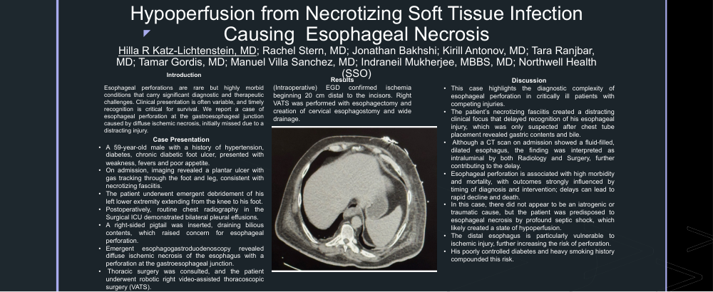
Discussion: This case highlights the diagnostic complexity of esophageal perforation in critically ill patients with competing injuries. The patient’s necrotizing fasciitis created a distracting clinical focus that delayed recognition of his esophageal injury, which was only suspected after chest tube placement revealed gastric contents and bile. Although a CT scan on admission showed a fluid-filled, dilated esophagus, the finding was interpreted as intraluminal by both Radiology and Surgery, further contributing to the delay. Esophageal perforation is associated with high morbidity and mortality, with outcomes strongly influenced by timing of diagnosis and intervention; delays, especially in unstable patients, can lead to rapid decline and death. In this case, there did not appear to be an iatrogenic or traumatic cause, but the patient was predisposed to esophageal necrosis by profound septic shock, which likely created a state of hypoperfusion. The distal esophagus is particularly vulnerable to ischemic injury, further increasing the risk of perforation. His poorly controlled diabetes and heavy smoking history compounded this risk.