119 posters, 6 topics, 524 authors, 243 institutions
ePostersLive by SciGen Technologies S.A. All rights reserved.
RCR Global AI Conference 2026
29-30 June, 2026 | QEII Centre, Westminster

95
A National Evaluation of UK Radiology Resident Experiences with Artificial Intelligence-Derived Acute Stroke Imaging Software
Poster Presenter
Authors
Timothy Cheng, Jake Cowen, Jason Mak, Maurizio Parker, RADIANT Collaborative Group, Adrian Tang, Yogish Joshi, Helen Addley, Thomas Booth, Fiona Gilbert, Abhishekh Ashok
Affiliations
Department of Radiology, Medway Maritime Hospital, Medway NHS Foundation Trust, Gillingham ME7 5NY, Department of Radiology, Queen Alexandra Hospital, Portsmouth Hospitals University NHS Trust, PO6 3LY, United Kingdom., Department of Radiology, University College London Hospitals NHS Foundation Trust, 235 Euston Road, London, NW1 2BU, Radiology Academic Network For Trainees (RADIANT), Faculty of Biology, Medicine and Health, The University of Manchester, Department of Radiology, Cambridge University Hospitals NHS Foundation Trust, Cambridge CB2 0QQ, United Kingdom; Department of Radiology, University of Cambridge, Cambridge, UK, School of Biomedical Engineering & Imaging Sciences, King’s College London, London UK; Department of Neuroradiology, King’s College Hospital, London, UK.
Part of Topic
Transforming practice and leadership: pilot or test data on implementation of AI into clinical practice, clinical feedback or patient perspectives
A National Evaluation of UK Radiology Resident Experiences with Artificial Intelligence-Derived Acute Stroke Imaging Software
Timothy Cheng1, Jake Cowen2, Jason Mak3, Maurizio Parker4, RADIANT Collaborative Group, Adrian Tang5, Yogish Joshi6,7, Helen Addley6,7, Fiona Gilbert6,7,Thomas Booth8,9, Abhishekh H Ashok6,7
1Department of Radiology, Medway Maritime Hospital, Gillingham, UK | 2Department of Radiology, Queen Alexandra Hospital, Portsmouth, UK | 3Department of Radiology, University College London Hospitals NHS Foundation Trust, London, UK | 4Department of Radiology, Royal Sussex County Hospital, Brighton, UK | 5Faculty of Biology, Medicine and Health, The University of Manchester, UK | 6Department of Radiology, Cambridge University Hospitals NHS Foundation Trust, Cambridge, UK | 7Department of Radiology, University of Cambridge, Cambridge, UK | 8School of Biomedical Engineering & Imaging Sciences, King’s College London, London, UK | 9Department of Neuroradiology, King’s College Hospital, London, UK.
RADIANT Collaborative Group: Mohammad Abu-Hantasha, Hannah Adamsb, Ali Forookhic, Lara Jehanlid, Islam Noamane, Tom Poundallf, Natalie Sanzoneg, Hani Wahabh
aDepartment of Radiology, Queen Alexandra Hospital, Portsmouth, UK | bDepartment of Radiology, East Sussex Healthcare NHS Trust, Sussex, UK | cDepartment of Radiology, Norfolk and Norwich University Hospital, Norwich, UK | dDepartment of Radiology, Mersey and West Lancashire Teaching Hospitals, Prescot, UK | eDepartment of Radiology, Royal Infirmary of Edinburgh, Edinburgh, UK | fDepartment of Radiology, Nottingham University Hospitals NHS Trust, Nottingham, UK | gDepartment of Radiology, NHS Greater Glasgow and Clyde, Glasgow, UK | hDepartment of Radiology, Royal Victoria Infirmary, Newcastle Hospitals NHS Foundation Trust, Newcastle, UK.
Introduction
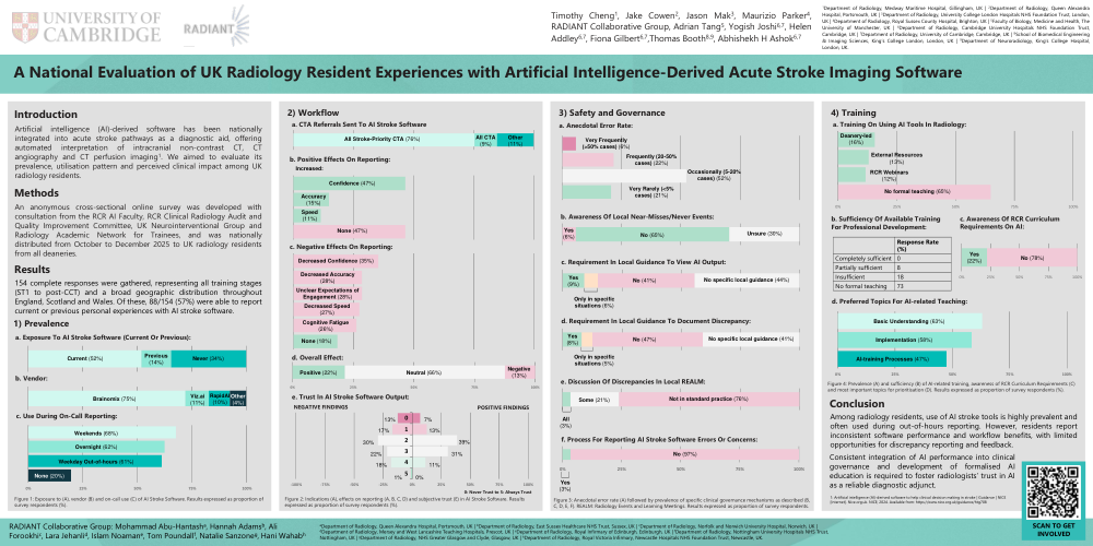
Artificial intelligence (AI)-derived software has been nationally integrated into acute stroke pathways as a diagnostic aid, offering automated interpretation of intracranial non-contrast CT, CT angiography and CT perfusion imaging1. We aimed to evaluate its prevalence, utilisation pattern and perceived clinical impact among UK radiology residents.
Methods
An anonymous cross-sectional online survey was developed with consultation from the RCR AI Faculty, RCR Clinical Radiology Audit and Quality Improvement Committee, UK Neurointerventional Group and Radiology Academic Network for Trainees, and was nationally distributed from October to December 2025 to UK radiology residents from all deaneries.
Results
154 complete responses were gathered, representing all training stages (ST1 to post-CCT) and a broad geographic distribution throughout England, Scotland and Wales. Of these, 88/154 (57%) were able to report current or previous personal experiences with AI stroke software.
1) Prevalence
a. Exposure To AI Stroke Software (Current Or Previous): Current: 52%; Previous: 14%; Never: 34%
b. Vendor: Brainomix: 75%; Viz.ai: 11%; RapidAI: 10%; Other: 4%
c. Use During On-Call Reporting: Weekends: 68%; Overnight: 62%; Weekday Out-of-hours: 61%; None: 20%
Figure 1 Caption: Figure 1: Exposure to (A), vendor (B) and on-call use (C) of AI Stroke Software. Results expressed as proportion of survey respondents (%).
2) Workflow
a. CTA Referrals Sent To AI Stroke Software: All Stroke-Priority CTA: 76%; All CTA: 9%; Other: 11%
b. Positive Effects On Reporting (Increased): Confidence: 47%; Accuracy: 15%; Speed: 11%; None: 47%
c. Negative Effects On Reporting: Decreased Confidence: 35%; Decreased Accuracy: 28%; Unclear Expectations of Engagement: 28%; Decreased Speed: 27%; Cognitive Fatigue: 26%; None: 18%
d. Overall Effect: Positive: 22%; Neutral: 66%; Negative: 13%
e. Trust In AI Stroke Software Output (0: Never Trust to 5: Always Trust):
0: Negative Findings (13%), Positive Findings (7%)
1: Negative Findings (17%), Positive Findings (13%)
2: Negative Findings (30%), Positive Findings (39%)
3: Negative Findings (22%), Positive Findings (31%)
4: Negative Findings (18%), Positive Findings (11%)
5: Negative Findings (1%), Positive Findings (0%)
Figure 2 Caption: Figure 2: Indications (A), effects on reporting (A, B, C, D) and subjective trust (E) in AI Stroke Software. Results expressed as proportion of survey respondents (%).
3) Safety and Governance
a. Anecdotal Error Rate: Very Frequently (>50% cases): 6%; Frequently (20-50% cases): 22%; Occasionally (5-20% cases): 52%; Very Rarely (<5% cases): 21%
b. Awareness Of Local Near-Misses/Never Events: Yes: 6%; No: 65%; Unsure: 30%
c. Requirement In Local Guidance To View AI Output: Yes: 9%; Only in specific situations: 6%; No: 41%; No specific local guidance: 44%
d. Requirement In Local Guidance To Document Discrepancy: Yes: 8%; Only in specific situations: 5%; No: 47%; No specific local guidance: 41%
e. Discussion Of Discrepancies In Local REALM: All: 3%; Some: 21%; Not in standard practice: 76%
f. Process For Reporting AI Stroke Software Errors Or Concerns: Yes: 3%; No: 97%
Figure 3 Caption: Figure 3: Anecdotal error rate (A) followed by prevalence of specific clinical governance mechanisms as described (B, C, D, E, F). REALM: Radiology Events and Learning Meetings. Results expressed as proportion of survey respondents.
4) Training
a. Training On Using AI Tools In Radiology: Deanery-led: 16%; External Resources: 13%; RCR Webinars: 12%; No formal teaching: 65%
b. Sufficiency Of Available Training For Professional Development: Completely sufficient: 0%; Partially sufficient: 8%; Insufficient: 18%; No formal teaching 73%
c. Awareness Of RCR Curriculum Requirements On AI: Yes: 22%; No: 78%
d. Preferred Topics For AI-related Teaching: Basic Understanding: 63%; Implementation: 58%; AI-training Processes: 47%
Figure 4 Caption: Figure 4: Prevalence (A) and sufficiency (B) of AI-related training, awareness of RCR Curriculum Requirements (C) and most important topics for prioritisation (D). Results expressed as proportion of survey respondents (%).
Conclusion
Among radiology residents, use of AI stroke tools is highly prevalent and often used during out-of-hours reporting. However, residents report inconsistent software performance and workflow benefits, with limited opportunities for discrepancy reporting and feedback.
Consistent integration of AI performance into clinical governance and development of formalised AI education is required to foster radiologists’ trust in AI as a reliable diagnostic adjunct.