119 posters, 6 topics, 524 authors, 243 institutions
ePostersLive by SciGen Technologies S.A. All rights reserved.
RCR Global AI Conference 2026
29-30 June, 2026 | QEII Centre, Westminster

64
Radiology Service Delivery in the Future: Which "Toilsome" aspects of their current role would Radiologists wish to delegate to an AI Agent ? Design of a baseline audit of Radiologist and Referring Clinicians' Views
Part of Topic
Transforming practice and leadership: pilot or test data on implementation of AI into clinical practice, clinical feedback or patient perspectives
TITLE
The Future of Agentic AI in Radiology: A Reaudit of Radiologist and Referrer Views
AFFILIATIONS
University of Manchester, Manchester, UK.
Queen’s Hospital, BHRUT (Teaching Hospital Trust), Romford, UK.
Manchester University NHS Foundation Trust, UK.
BACKGROUND
Current AI tools in Radiology are static, task-specific applications with little to no access to clinical context (electronic patient records - EPR) or prior imaging (1).
The conceptualization of agentic AI systems has risen in recent medical literature (2,3).
Agentic AI is defined as a system capable of autonomous task execution, advanced reasoning, adaptive decision-making, and dynamic human-AI interaction.
Baseline audit data emphasizes the need to encourage radiologists to influence the development and certification of useful rather than easily marketable AI (4).
COMPARING CURRENT AI VS. AGENTIC AI
Current Radiology AI: Finely tuned CNN trained on large imaging datasets. Vulnerable to out-of-distribution/data drift, mandating intensive and expensive post-market surveillance.
Proposed Agentic AI: Multi-agent entity orchestrating agents trained on various models. Less vulnerable to any single mode of AI failure.
AIMS
To define the concept of an agentic AI within radiology: an "Apprentice Agent" working under a "Master Radiologist".
To assess conceptual understanding amongst clinicians on the proposed AI model.
To reaudit the willingness of radiologists and referrers to delegate a variety of clinical and non-clinical tasks to Agent R following the optimization of the data collection tool.
INTRODUCING AGENT R
A hypothetical autonomous AI entity unique to and trainable by each Radiologist.
Fully EPR Aware
Chat & Speech Functionality
Can say "I don't know" and seek supervision
Adaptive learning capabilities
METHODS
Two distinct surveys (Radiologist & Referrer) were designed using Microsoft Forms.
Distribution was completed via physical posters, QR codes, survey links within departments, mailing lists, and Grand Rounds. Participation was voluntary and anonymous.
Scoring was based on a Likert scale (0 - Not at all Likely, 10 - Very Likely).
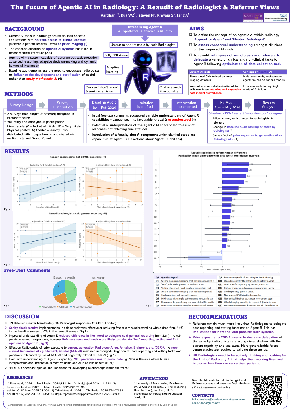
Initial free-text comments from the baseline audit suggested a variable understanding of Agent R capabilities, categorized into favorable, critical, and misunderstood (4).
Potential misinterpretation of the agentic AI concept led to a risk of responses not reflecting true attitudes.
To resolve this, a "sanity check" component was introduced consisting of 3 questions clarifying the scope and capabilities of Agent R.
Success Criterion: Less than 10% free-text responses falling into the "misunderstood" category.
Figure 1 multivariate regression was performed by Copilot @ MFT.
RESULTS & DISCUSSION
Sample Size: 19 Referrer responses (Greater Manchester), 16 Radiologist responses (13 Greater Manchester, 3 London).
Sanity Check Results: Implementation in this re-audit was effective at reducing free-text misunderstanding, showing a drop from 31% in the baseline survey to 0% in the re-audit survey (Fig 2).
Closing the Gap on Cold Reporting: Improved understanding of Agent R reduced the difference in likelihood to delegate cold general reporting from 3.8 points (4) to 0.5 points in re-audit responders.
Hot Reporting & Vetting: Referrers remained much more likely than radiologists to delegate "hot" reporting/vetting and 2nd opinions to Agent R (Fig 3).
Effect of Prior AI Exposure: The impact of prior technology exposure on radiologists remained unchanged. Delegation of core reporting and vetting tasks was positively influenced by the use of Non-Clinical Generative AI (NCG-AI like ChatGPT, Copilot) and negatively related to prior use of current clinical Radiology AI (CGR-AI like Annalise, Brainomix) (Fig 1).
The Value of MDTs: Even with a clear understanding of Agent R capabilities, the strong preference was for human participation in Multidisciplinary Teams (MDTs).
Quote 1: "This is the area where human interpretation and interaction is most valuable and AI is of less benefit (MDT)."
Quote 2: "MDT is a specialist opinion and important for developing relationships within the team."
RECOMMENDATIONS
Procurement Implications: Referrers remain much more likely than Radiologists to delegate core reporting and vetting functions to Agent R. This has major implications for how and who procures such systems.
Address Current Dissatisfaction: Prior exposure to current-generation radiology AI (CGR-AI) seems to decrease task delegation by Radiologists, suggesting dissatisfaction with current capabilities and use cases. More generalizable/cross-centre studies are required to validate these trends.
Active Professional Influence: UK Radiologists need to be actively thinking and pushing for the specific kind of Radiology AI that actually helps their working lives and improves how they serve patients.
ADMINISTRATIVE INFO
References:
Katal et al., 2024 — Eur J Radiol. 2024;181. doi:10.1016/j.ejrad.2024.111798.
Karunanayake et al., 2025 — Inform Health. 2025;2(2):73–83. doi:10.1016/j.infoh.2025.03.001.
Chinniah et al., 2026 — Clin Radiol. 2026;97:107351. doi:10.1016/j.crad.2026.107351.
Baseline Audit Findings — https://epos.myesr.org/poster/esr/ecr2026/C-28903
Contacts:
inika.vardhan@student.manchester.ac.uk
adrian.tang@nhs.net
External Links:
Scan the QR code or go to links.tangoscan.com/rcrAI for full Radiologist and Referrer surveys and baseline Audit findings.
Note:
Concept image of Agent R by OpenAI from an author-defined prompt; used for illustrative purposes only.
Figures referenced: Fig 1a, Fig 1b, Fig 2, Fig 3.