119 posters, 6 topics, 524 authors, 243 institutions
ePostersLive by SciGen Technologies S.A. All rights reserved.
RCR Global AI Conference 2026
29-30 June, 2026 | QEII Centre, Westminster

134
Mind the Gap: Assessing the Generalizability of a Public AI Model for Intracranial Hemorrhage Detection on Non-Contrast CT
Poster Presenter
Authors
Rabia Shaukat, Ali Mansoor, Fatima Iqbal, Muhammad Arslan, Muneeb Ahmad, Nighat Haroon Khan
Affiliations
Lahore General Hospital, Eastern Medical Technology Services, Lahore General Hospital / Ameer ud Din Medical College/ Post Graduate Medical Institute, Lahore General Hospital / Ameer ud Din Medical College / Post Graduate Medical Institute, Lahore General Hospital/ Ameer ud Din Medical College / Post Graduate Medical Institute, Jinnah Hospital Lahore
Part of Topic
Transforming practice and leadership: pilot or test data on implementation of AI into clinical practice, clinical feedback or patient perspectives
Background:
Spontaneous intracranial haemorrhage (ICH) carries high mortality (30–50%) and significant long-term disability (1,2). In low- and middle-income countries such as Pakistan, delayed diagnosis is worsened by limited access to expert imaging interpretation (3). Early detection on non-contrast CT is therefore critical. Artificial intelligence (AI), particularly deep learning models, has shown strong performance in ICH detection, with reported AUCs of 0.85–0.96 (4). However, approved AI tools remain limited in LMIC settings, and real-world comparative data against clinical readers in Pakistan are scarce.
Objective:
To compare the diagnostic accuracy of an AI algorithm with radiologists of varying experience levels in detecting acute ICH on non-contrast CT scans.
Materials and Methods:
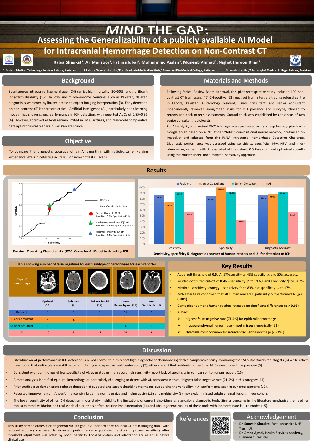
Following Ethical Review Board approval, this pilot retrospective study included 100 non-contrast CT brain scans (47 ICH-positive, 53 negative) from a tertiary trauma referral centre in Lahore, Pakistan. A radiology resident, junior consultant, and senior consultant independently reviewed anonymized scans for ICH presence and subtype, blinded to reports and each other’s assessments. Ground truth was established by consensus of two senior consultant radiologists.For AI analysis, anonymized DICOM images were processed using a deep learning pipeline in Google Colab based on a 2D EfficientNet-B3 convolutional neural network, pretrained on ImageNet and adapted from the RSNA Intracranial Hemorrhage Detection Challenge. Diagnostic performance was assessed using sensitivity, specificity, PPV, NPV, and inter-observer agreement, with AI evaluated at the default 0.5 threshold and optimized cut-offs using the Youden Index and a maximal-sensitivity approach.
Key Results:
This study demonstrates a clear generalizability gap in AI performance on local CT brain imaging data, with reduced accuracy compared to expected performance in published settings. Improved sensitivity after threshold adjustment was offset by poor specificity. Local validation and adaptation are essential before clinical use.