1,830 posters, 67 videos, 75 audios, 29 topics, 22 sessions, 4,727 authors, 1,697 institutions
ePostersLive by SciGen Technologies S.A. All rights reserved.
RCOG World Congress 2023
June 12 - 14, 2023 | London, UK

EP.1411
Life-threatening Hemoperitoneum and Sepsis with Giant abdominal mass of diagnostic uncertainty.
Poster Presenter
Authors
Affiliations
Part of Topic
Imaging in Gynaecology
Life-threatening Hemoperitoneum and Sepsis with Giant abdominal mass of diagnostic uncertainty
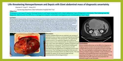
Objective: To present a rare case of massive Hemoperitoneum and Sepsis associated with a Giant abdominal mass of diagnostic uncertainty.The challenge was the diagnostic dilemma and the difficulty in management despite multimodal investigations and multidisciplinary team input.
Case Report: 58-years old post-menopausal lady, with history of fibroid presented to the emergency department with acute abdominal pain, distension and vomiting. She was pale, unwell and had a vasovagal syncope. Her CT showed a large mass of 20 x 20 x15 cm with Omental stranding and Ascites. She became hypotensive overnight with a significant drop of haemoglobin and developed sepsis. Ultrasound showed haemoperitoneum, and a large multilocular solid-cystic complex mass. Uterus was clearly visualised with no fibroid, normal left ovary and right ovary was not visualised. Her CA-125 was 302. Patient was transfused and received broad spectrum antibiotics. She had an MRI which was reviewed in the joint radiologist-gynaecologist meeting, where it was concluded that the large mass is either a benign fibroid with haemorrhage and degeneration or a right ovarian mass with intermittent torsion. She continued to detoriateclinically and was counselled for surgery. In view of high suspicion for ovarian malignancy she had a midline laparotomy with full staging procedure. There wasa 25 cm malignant looking ovarian mass on the right side with haemorrhagic spillage. There was one litre of haemoperitoneumin the Pelvis. She received further blood transfusion and made a good recovery. Her histology confirms Benign Serous Cystadenofibromawith partial infarction.
Discussion: Ovarian Cystadenofibromasare relatively rare variants of epithelial tumours of ovary containing epithelial and fibrous stromal components. They account for 1.7% of all benign ovarian tumours. Pre-operative diagnosis is difficult as these tumours mimic characteresticsof malignancy such as large cystic mass with septations, papillary projections and solid nodules on ultrasound. The Omental stranding and ascites was also misleading. These tumours can also resemble the gross appearance of malignant tumours peri-operatively. Final histology showed partial infarction and intravascular thrombosis which can reflect torsion. This was possibly the reason for the haemorrhage and was misinterpreted as fibroid degeneration.
Conclusions: Serous cystadenofibromascan be difficult to diagnose on imaging. Ovarian torsion can be associated with serous cystadenofibromasdue to its tendency to grow into large size. This case highlights multidisciplinary approach and utilising multiple imaging modalities. It also emphasize the importance of a detailed discussion with the patient regarding the findings on the scans and the nature of mass. Careful planning, MDT approach and preparation for extensive surgery is also advisable.