58 posters, 14 topics, 4 sessions, 188 authors, 62 institutions
ePostersLive by SciGen Technologies S.A. All rights reserved.
PSRC 69th Annual Meeting 2024
May 16-19, 2024 | The Westin Copley Place, Boston, MA

EP04
Understanding How Silicone Breast Implant Shells Reduce Proliferation Rates Of Both Benign And Malignant Cell Lines
Poster Presenter
Authors
Affiliations
Part of Topic
Breast
Understanding How Silicone Breast Implant Shells Reduce Proliferation Rates of both Benign and Malignant Cell Lines
Jini Jeon BA, Riley Mayne BS, Tim Y. Li BA BS, Dylan K. Kim AB, Xue Dong MD PhD, and Jason A. Spector MD FACS
INTRODUCTION
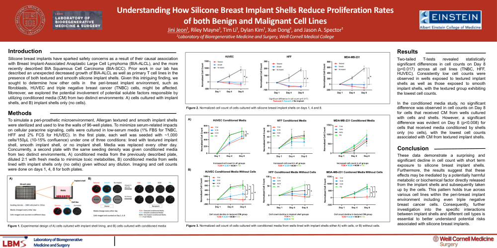
Silicone breast implants have sparked safety concerns as a result of their causal association with Breast Implant-Associated Anaplastic Large Cell Lymphoma (BIA-ALCL), and the more recently described BIA Squamous Cell Carcinoma (BIA-SCC). Prior work in our lab has described an unexpected decreased growth of BIA-ALCL as well as primary T cell lines in the presence of both textured and smooth silicone implant shells. Given this intriguing finding, we sought to determine how other cells in the peri-breast implant environment, such as fibroblasts, HUVEC and triple negative breast cancer (TNBC) cells, might be affected. Moreover, we explored the potential involvement of potential soluble factors responsible by utilizing conditioned media (CM) from two distinct environments: A) cells cultured with implant shells, and B) implant shells only (no cells).
METHODS
To simulate a peri-prosthetic microenvironment, Allergan textured and smooth implant shells were sterilized and used to line the walls of 96-well plates. To minimize serum-related impacts on cellular paracrine signaling, cells were cultured in low-serum media (1% FBS for TNBC, HFF and 2% FCS for HUVEC). In the first plate, each well was seeded with ~1,000 cells/150µL (10-15% confluence) under one of three conditions: lined with textured implant shell, smooth implant shell, or no implant shell. Media was replaced every other day. Concurrently, a second plate with the same seeding density was given conditioned media from two distinct environments, A) conditioned media from the previously described plate, diluted 2:1 with fresh media to minimize toxic metabolites, B) conditioned media from wells lined with implant shells only (no cells) given without any dilution. Imaging and cell counts were done on days 1, 4, 8 for both plates.
RESULTS
Two-tailed T-tests revealed statistically significant differences in cell counts on Day 8 (p<0.017) across all cell lines (TNBC, HFF, HUVEC). Consistently low cell counts were observed in wells exposed to textured implant shells as well as those exposed to smooth implant shells, with the textured group exhibiting the lowest cell counts. In the conditioned media study, no significant difference was observed in cell counts on Day 8 for cells that received CM from wells cultured with cells and shells. However, a significant difference was evident on Day 8 (p<0.008) for cells that received media conditioned by shells only (no cells), with the lowest cell counts associated with CM from textured implant shells.
CONCLUSION
These data demonstrate a surprising and significant decline in cell count with short term exposure to silicone breast implant shells. Furthermore, the results suggest that these effects may be mediated by a potentially harmful metabolic or biochemical factor directly released from the implant shells and subsequently taken up by the cells. This pattern holds true across various cell lines within the peri-breast implant environment including even triple negative breast cancer cells. Consequently, further investigation into the specific interactions between implant shells and different cell types is essential to better understand potential risks associated with silicone breast implants.