58 posters, 14 topics, 4 sessions, 188 authors, 62 institutions
ePostersLive by SciGen Technologies S.A. All rights reserved.
PSRC 69th Annual Meeting 2024
May 16-19, 2024 | The Westin Copley Place, Boston, MA

EP41
Development Of Tissue Adhesives For Sutureless Microvascular Anastomosis
Poster Presenter
Part of Topic
Microsurgery/Flap Physiology/Ischemia Reperfusion
Development Of Tissue Adhesives For Sutureless Microvascular Anastomosis
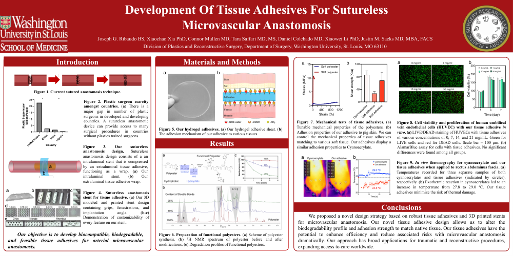
Figure 2. Plastic surgeon scarcity amongst countries. (a) There is a major gap in number of plastic surgeons in developed and developing countries. A sutureless anastomotic device can provide access to many surgical procedures in countries without plastics trained surgeons.
Figure 3. Our sutureless anastomosis design. Sutureless anastomosis design consists of a an intralumenal stent that is compressed by an extraluminal tissue adhesive, functioning as a wrap. (a) Our intraluminal stent. (b) Our extraluminal tissue adhesive wrap.
Figure 4. Sutureless anastomosis stent for tissue adhesive. (a) Our 3D modeled and printed stent design containing grips, fenestrations, and implantation angle. (b-e) Demonstration of customizability of every feature on our stent.
Our objective is to develop biocompatible, biodegradable, and feasible tissue adhesives for arterial microvascular anastomosis.
Figure 5. Our hydrogel adhesives. (a) Our hydrogel adhesive sheet. (b) The adhesion mechanism of our adhesive to various tissues.
Figure 6. Preparation of functional polyesters. (a) Scheme of polyester synthesis. (b) 1H NMR spectrum of polyester before and after modifications. (c) Degradation profiles of functional polyesters.
Figure 7. Mechanical tests of tissue adhesives. (a) Tunable mechanical properties of the polyesters. (b) Adhesion properties of our adhesive to pig skin. We can control the mechanical properties of tissue adhesives matching to various soft tissue. Our adhesives display a similar adhesion properties to Cyanoacrylate.
Figure 8. Cell viability and proliferation of human umbilical vein endothelial cells (HUVEC) with our tissue adhesive in vitro. (a) LIVE/DEAD staining of HUVECs with tissue adhesives of various concentrations of 0, 7, 14, and 21 mg/mL. Green for LIVE cells and red for DEAD cells. Scale bar = 100 μm. (b) AlamarBlue assay for cells with tissue adhesives. No significant differences were found among all groups.
Figure 9. In vivo thermography for cyanoacrylate and our tissue adhesives when applied to rectus abdominus fascia. (a) Temperatures recorded for three separate samples of both cyanoacrylates and tissue adhesives (indicated by circles), respectively. (b) Exothermic reaction in cyanoacrylates led to an increase in temperature from 27.8 to 29.0 ºC. Our tissue adhesives minimize the risk of thermal damage.
We proposed a novel design strategy based on robust tissue adhesives and 3D printed stents for microvascular anastomosis. Our novel tissue adhesive design allows us to alter the biodegradability profile and adhesion strength to match native tissue. Our tissue adhesives have the potential to enhance efficiency and reduce associated risks with microvascular anastomosis dramatically. Our approach has broad applications for traumatic and reconstructive procedures, expanding access to care worldwide.