358 posters, 97 topics, 122 sessions, 958 authors, 239 institutions
ePostersLive by SciGen Technologies S.A. All rights reserved.
79th PostGraduate Assembly in Anesthesiology 2025
December 12 - 15, 2025 | New York City

2126
Hemiazygous Vein Trialysis Cannulation Falsely Appearing as Extravascular Mediastinal Placement after Liver Transplant. A Case Report.
Poster Presenter
Part of Topic
Cardiovascular - Transplant
Introductiton
Central venous catheters are essential for administering medications that can damage peripheral veins (e.g., vasopressors, chemotherapeutic agents, hypertonic solutions), providing long-term intravenous therapy, enabling hemodynamic monitoring, and facilitating hemodialysis or plasmapheresis. They are also invaluable when peripheral venous access is difficult or impossible.
Although central line placement is a common procedure, it carries the risk of serious complications, including inadvertent cannulation of the azygos vein via the internal jugular vein (IJV). The azygos vein is typically small but may become dilated in certain pathological conditions, increasing the likelihood of accidental cannulation.
Case presentation and clinical course
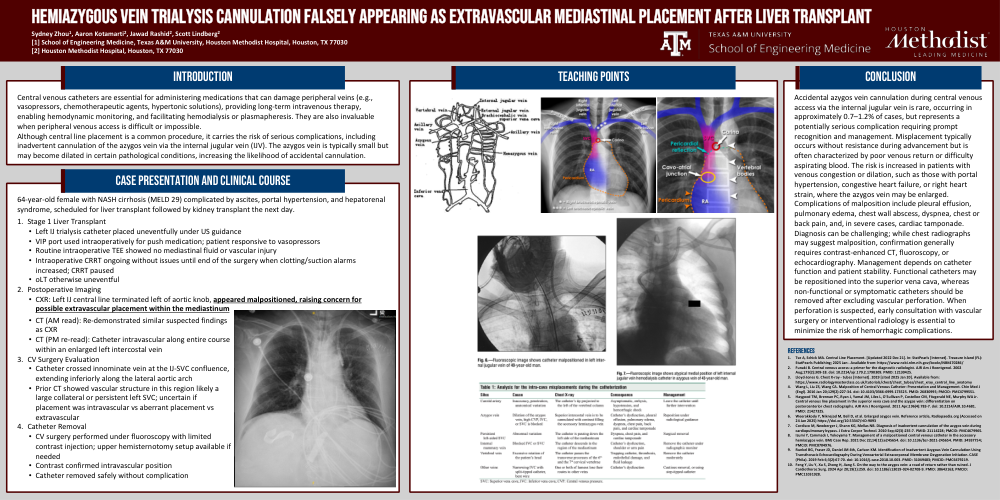
Accidental azygos vein cannulation during central venous access via the internal jugular vein is rare, occurring in approximately 0.7–1.2% of cases, but represents a potentially serious complication requiring prompt recognition and management. Misplacement typically occurs without resistance during advancement but is often characterized by poor venous return or difficulty aspirating blood. The risk is increased in patients with venous congestion or dilation, such as those with portal hypertension, congestive heart failure, or right heart strain, where the azygos vein may be enlarged. Complications of malposition include pleural effusion, pulmonary edema, chest wall abscess, dyspnea, chest or back pain, and, in severe cases, cardiac tamponade. Diagnosis can be challenging; while chest radiographs may suggest malposition, confirmation generally requires contrast-enhanced CT, fluoroscopy, or echocardiography. Management depends on catheter function and patient stability. Functional catheters may be repositioned into the superior vena cava, whereas non-functional or symptomatic catheters should be removed after excluding vascular perforation. When perforation is suspected, early consultation with vascular surgery or interventional radiology is essential to minimize the risk of hemorrhagic complications.