358 posters, 97 topics, 122 sessions, 958 authors, 239 institutions
ePostersLive by SciGen Technologies S.A. All rights reserved.
79th PostGraduate Assembly in Anesthesiology 2025
December 12 - 15, 2025 | New York City

2205
Intraoperative Identification of a Right Ventricular Blood Cyst
Part of Topic
Cardiovascular
| Intraoperative Identification of a Right Ventricular Blood Cyst | |
| This case describes the anesthesiologist's role in the intraoperative diagnosis and management of unexpected cardiac masses, with particular emphasis on the utility of transesophageal echocardiography (TEE) for real-time decision making. | |
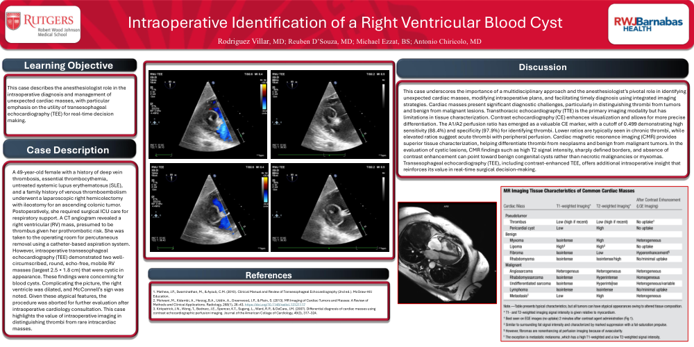
| A 49-year-old female with a history of deep vein thrombosis, essential thrombocythemia, untreated systemic lupus erythematosus (SLE), and a family history of venous thromboembolism underwent a laparoscopic right hemicolectomy with ileostomy for an ascending colonic tumor. Postoperatively, she required surgical ICU care for respiratory support. A CT angiogram revealed a right ventricular (RV) mass, presumed to be thrombus given her prothrombotic risk. She was taken to the operating room for percutaneous removal using a catheter-based aspiration system. However, intraoperative transesophageal echocardiography (TEE) demonstrated two well-circumscribed, round, echo-free, mobile RV masses (largest 2.5 × 1.8 cm) that were cystic in appearance. These findings were concerning for blood cysts. Complicating the picture, the right ventricle was dilated, and McConnell’s sign was noted. Given these atypical features, the procedure was aborted for further evaluation after intraoperative cardiology consultation. This case highlights the value of intraoperative imaging in distinguishing thrombi from rare intracardiac masses. | |
| This case underscores the importance of a multidisciplinary approach and the anesthesiologist’s pivotal role in identifying unexpected cardiac masses, modifying intraoperative plans, and facilitating timely diagnosis using integrated imaging strategies. Cardiac masses present significant diagnostic challenges, particularly in distinguishing thrombi from tumors and benign from malignant lesions. Transthoracic echocardiography (TTE) is the primary imaging modality but has limitations in tissue characterization. Contrast echocardiography (CE) enhances visualization and allows for more precise differentiation. The A1/A2 perfusion ratio has emerged as a valuable CE marker, with a cutoff of 0.499 demonstrating high sensitivity (88.4%) and specificity (97.9%) for identifying thrombi. Lower ratios are typically seen in chronic thrombi, while elevated ratios suggest acute thrombi with peripheral perfusion. Cardiac magnetic resonance imaging (CMR) provides superior tissue characterization, helping differentiate thrombi from neoplasms and benign from malignant tumors. In the evaluation of cystic lesions, CMR findings such as high T2 signal intensity, sharply defined borders, and absence of contrast enhancement can point toward benign congenital cysts rather than necrotic malignancies or myxomas. Transesophageal echocardiography (TEE), including contrast-enhanced TEE, offers additional intraoperative insight that reinforces its value in real-time surgical decision-making. We plan to present the Echo loops during our presentation. |
|
| 1. Mathew, J.P., Swaminathan, M., & Ayoub, C.M. (2010). Clinical Manual and Review of Transesophageal Echocardiography (2nd ed.). McGraw-Hill Education. 2. Motwani, M., Kidambi, A., Herzog, B.A., Uddin, A., Greenwood, J.P., & Plein, S. (2013). MR Imaging of Cardiac Tumors and Masses: A Review of Methods and Clinical Applications. Radiology, 268(1), 26–43. https://doi.org/10.1148/radiol.13121117 3. Kirkpatrick, J.N., Wong, T., Bednarz, J.E., Spencer, K.T., Sugeng, L., Ward, R.P., & DeCara, J.M. (2007). Differential diagnosis of cardiac masses using contrast echocardiographic perfusion imaging. Journal of the American College of Cardiology, 49(3), 317–324. |