358 posters, 97 topics, 122 sessions, 958 authors, 239 institutions
ePostersLive by SciGen Technologies S.A. All rights reserved.
79th PostGraduate Assembly in Anesthesiology 2025
December 12 - 15, 2025 | New York City

2044
Percutaneous Ventricular Assist Devices as a Perioperative Lifeline: Anesthetic Management for Severe Cardiomyopathy in High-Risk Colorectal Surgery
Poster Presenter
Authors
Eugene Michanine, Rafael Cabrales, Jamshed Desai, Alfredo Terrero Rodriguez
Affiliations
Part of Topic
Cardiovascular - Perioperative Management
Percutaneous Ventricular Assist Devices as a Perioperative Lifeline: Anesthetic Management for Severe Cardiomyopathy in High-Risk Colorectal Surgery
Michanine, E., Cabrales, R., Desai, J., & Rodriguez, A. T.
Department of Anesthesiology, Cleveland Clinic Florida
Background
Patients with severe systolic heart failure undergoing major noncardiac surgery face high perioperative mortality due to hemodynamic instability, poor contractility, and limited physiologic reserve. Standard strategies, such as inotropes and balloon pumps, may be inadequate for major non-cardiac surgeries.
Temporary percutaneous ventricular assist devices (pVADs) such as the Impella 5.5 provide left ventricle (LV) unloading and up to 4–5 L/min of forward flow, enabling “protected” noncardiac operations for patients otherwise considered inoperable.
This case reports intraoperative Impella 5.5 placement to facilitate urgent colorectal resection in a patient with end-stage nonischemic cardiomyopathy, highlighting anesthetic strategy, hemodynamic management, and multidisciplinary coordination.
Case Presentation
Patient Characteristics
66-year-old male with hypertension, obstructive sleep apnea, Barrett’s esophagus, and T2 rectal cancer treated with chemoradiation (5-FU & methotrexate), nonischemic cardiomyopathy (EF 13–21%), ICD, CardioMEMS, NYHA III symptoms, and progressive cardiogenic shock (SCAI D), was admitted for dyspnea and edema. RHC revealed volume overload (CO 2.6, CI 1.1) and LHC showed nonocclusive disease in LAD and LCx, occlusive in RCA. He initially required aggressive diuresis, milrinone at 0.25 mcg/kg/min, and femoral converted to left axillary IABP. Worsening malignancy mandated urgent proctectomy; durable LVAD was deferred due to bleeding risk from anticoagulation.
Preop hemodynamics: BP 89/54, HR 77, PA pressures 54/33 mmHg, IABP 1:1 augmentation ~120%. Labs acceptable; Hgb 12.5, renal function preserved. ICD tachyarrhythmias were suspended and external defibrillator pads were applied.
Intraoperative Course
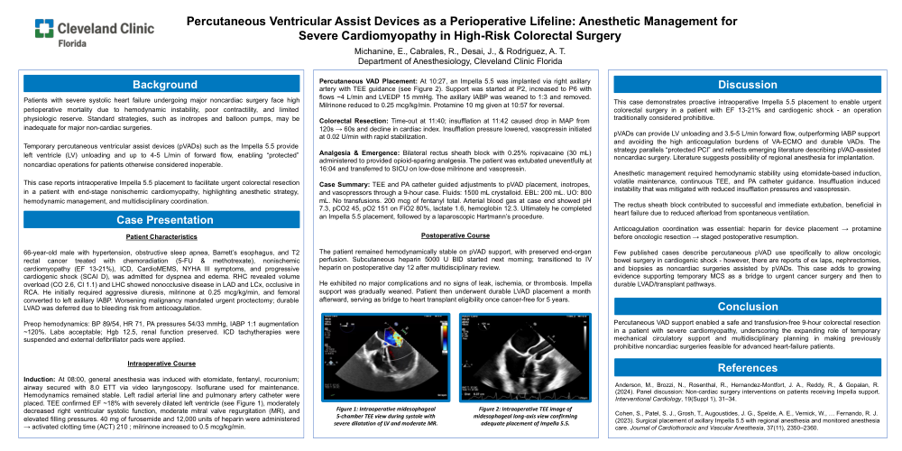
Induction: At 08:00, general anesthesia was induced with etomidate, fentanyl, rocuronium; airway secured with 8.0 ETT via video laryngoscopy. Isoflurane was used for maintenance. Hemodynamics remained stable. Left radial arterial line and right pulmonary artery catheter were placed. TEE confirmed EF ~18% with severely dilated left ventricle (see Figure 1), moderately decreased right ventricular systolic function, moderate mitral regurgitation (MR), and elevated filling pressures. 40 mg of furosemide and 12,000 units of heparin were administered → activated clotting time (ACT) 210; milrinone increased to 0.5 mcg/kg/min.
Percutaneous VAD Placement: At 10:27, an Impella 5.5 was implanted via right axillary artery with TEE guidance (see Figure 2). Support was started at P2, increased to P6 with flows ~4 L/min and LVEDP 15 mmHg. The axillary IABP was weaned to 1:3 and removed. Milrinone reduced to 0.25 mcg/kg/min. Protamine 10 mg given at 10:57 for reversal.
Colorectal Resection: Time-out at 11:40; insufflation at 11:42 caused drop in MAP from 120s → 60s and decline in cardiac index. Insufflation pressure lowered, vasopressin initiated at 0.02 U/min with rapid stabilization.
Analgesia & Emergence: Bilateral rectus sheath block with 0.25% ropivacaine (30 mL) administered to provide opioid-sparing analgesia. The patient was extubated uneventfully at 16:04 and transferred to SICU on low-dose milrinone and vasopressin.
Case Summary: TEE and PA catheter guided adjustments to pVAD placement, inotropes, and vasopressors through a 9-hour case. Fluids: 1500 mL crystalloid. EBL: 200 mL. UO: 800 mL. No transfusions. 200 mcg of fentanyl total. Arterial blood gas at case end showed pH 7.3, pCO2 45, pO2 151 on FiO2 80%, lactate 1.6, hemoglobin 12.3. Ultimately he completed an Impella 5.5 placement, followed by a laparoscopic Hartmann’s procedure.
Postoperative Course
The patient remained hemodynamically stable on pVAD support, with preserved end-organ perfusion. Subcutaneous heparin 5000 U BID started next morning; transitioned to IV heparin on postoperative day 12 after multidisciplinary review.
He exhibited no major complications and no signs of leak, ischemia, or thrombosis. Impella support was gradually weaned. Patient then underwent durable LVAD placement a month afterward, serving as bridge to heart transplant eligibility once cancer-free for 5 years.
Image Captions
Figure 1: Intraoperative midesophageal 5-chamber TEE view during systole with severe dilatation of LV and moderate MR.
Figure 2: Intraoperative TEE image of midesophageal long-axis view confirming adequate placement of Impella 5.5.
Discussion
This case demonstrates proactive intraoperative Impella 5.5 placement to enable urgent colorectal surgery in a patient with EF 13–21% and cardiogenic shock — an operation traditionally considered prohibitive.
pVADs can provide LV unloading and 3.5–5 L/min forward flow, outperforming IABP support and avoiding the high anticoagulation burdens of VA-ECMO and durable VADs. The strategy parallels “protected PCI” and reflects emerging literature describing pVAD-assisted noncardiac surgery. Literature suggests possibility of regional anesthesia for implantation.
Anesthetic management required hemodynamic stability using etomidate-based induction, volatile maintenance, continuous TEE, and PA catheter guidance. Insufflation induced instability that was mitigated with reduced insufflation pressures and vasopressin.
The rectus sheath block contributed to successful and immediate extubation, beneficial in heart failure due to reduced afterload from spontaneous ventilation.
Anticoagulation coordination was essential: heparin for device placement → protamine before oncologic resection → staged postoperative resumption.
Few published cases describe percutaneous pVAD use specifically to allow oncologic bowel surgery in cardiogenic shock — however, there are reports of ex laps, nephrectomies, and biopsies as noncardiac surgeries assisted by pVADs. This case adds to growing evidence supporting temporary MCS as a bridge to urgent cancer surgery and then to durable LVAD/transplant pathways.
Conclusion
Percutaneous VAD support enabled a safe and transfusion-free 9-hour colorectal resection in a patient with severe cardiomyopathy, underscoring the expanding role of temporary mechanical circulatory support and multidisciplinary planning in making previously prohibitive noncardiac surgeries feasible for advanced heart-failure patients.
References
-
Anderson, M., Brozzi, N., Rosenthal, R., Hernandez-Montfort, J. A., Reddy, R., & Gopalan, R. (2024). Panel discussion: Non-cardiac surgery interventions on patients receiving Impella support. Interventional Cardiology, 19(Suppl 1), 31–34.
-
Cohen, S., Patel, S. J., Grosh, T., Augustides, J. G., Spelde, A. E., Vernick, W., … Fernando, R. J. (2023). Surgical placement of axillary Impella 5.5 with regional anesthesia and monitored anesthesia care. Journal of Cardiothoracic and Vascular Anesthesia, 37(11), 2350–2360.