358 posters, 97 topics, 122 sessions, 958 authors, 239 institutions
ePostersLive by SciGen Technologies S.A. All rights reserved.
79th PostGraduate Assembly in Anesthesiology 2025
December 12 - 15, 2025 | New York City

3004
Distinct effects of sevoflurane and propofol on vascular permeability in vitro and in a mouse model
Poster Presenter
Authors
Affiliations
Part of Topic
Basic Research - Cardiovascular
barrier to maintain vascular integrity. General anesthetics comprise a
heterogeneous group of drugs with multiple functions and mechanisms of
action, which are not yet fully elucidated. It is suggested that general
anesthetics threaten endothelium stability by causing changes in vascular
tones. Sevoflurane has been shown to promote growth and proliferation of
human endothelial progenitor cells and thus may support tumor
metastasis by enhancing angiogenesis and tumor cell survival in hypoxic
environments. Different from inhaled sevoflurane, intravenous propofol
could the protection against oxidative stress in endothelial cells due to its
phenol-based structure as a free radical scavenger. However, for most
general anesthetics, its functional role on endothelium remains obscure.
Here we characterized the effects of commonly used general anesthetic
agents on mouse pulmonary endothelial cells (MPECs) in vitro and in
vivo and explored the potential targets.
RESULTS
The repair of pulmonary endothelial damage imposed by surgical
medicine such as general anesthetics and mechanical ventilation is
extremely important to patients. However, our understanding of
the cellular and molecular mechanisms that
regulate endothelial functions within the context of surgery are limited.
The precise molecular mechanisms underpinning these responses
remain to be elucidated and require renewed attention.
• RT-PCR and Western blot: RNA and proteins samples were extracted
from HUVEC/MPECs treated with different anesthetics or lung lysate
• ELISA analysis of VEGF from HUVEC/MPEC cells, mouse serum, and
lung lysate
• Immunofluorescent staining (IF): HIF 1α were stained in HUVEC/MPECs
• In vitro permeability assay: HUVEC/MPEC monolayers formed on PET
membranes were treated with sevoflurane or propofol. After treatments,
monolayer staining and FITC-Dextran permeability testing were
performed. Disruption of monolayer integrity by sevoflurane was
observable both visually and by quantification of fluorescence.
• Trans-endothelial electric resistance (TEER): : HUVEC/MPEC
monolayers formed on PET membranes were treated with sevoflurane
(1.3 MAC) or propofol (4 μg/mL) for three hours. After treatments, TEER
was measured to quantify the endothelium integrity.
• Rhodamine-dextran in vivo vascular permeability assay: In Balb/c mice,
one day after exposure to general anesthetics, rhodamine-dextran were
i.v. injected at 100 mg/kg. Three hours later, each mouse was
anesthetized and perfused with saline and followed by 5 mL of 4%
formaldehyde. Lung were cryosectioned and stained with Lycopersicon
Esculentum lectin. Vascular leakage was examined by fluorescence
microscope.
• AngioSense® 750 fluorescent in vivo vascular permeability assay: In
Balb/c mice, one day before exposure to general anesthetics,
AngioSense® 750 were i.v. injected. On the second day, mice were
exposure to sevoflurane or propofol for three hours and afterwards
mouse lung was harvested for Biofluorescent imaging using PerkinElmer
IVIS machine.
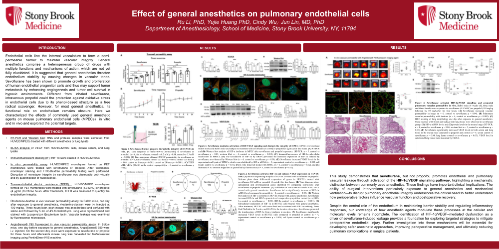
Figure 1. Sevoflurane but not propofol disrupts the integrity of
HUVECs in vitro. (A) Dose responses of trans-HUVEC
permeability to sevoflurane or propofol. (n = 6, for sevoflurane:
control vs 0.2 mM p =0.04, control vs 0.3 mM p =0.001). (B) Time
responses of trans-HUVEC permeability to sevoflurane or propofol.
(n = 6, for sevoflurane control vs 3-hour p = 0.046, control vs 4-
hour p = 0.03, control vs 6-hour p = 0.005, control vs 24-hour p <
0.0001). (C) TEER of HUVECs. (DMSO as the control to propofol)
(n = 6, control vs sevoflurane p = 0.02)
Figure 3. Sevoflurane mediates activation of HIF/VEGF signaling and disrupts the integrity of MPEC. MPECs were isolated from 6 weeks old
Balb/c mice and subject to treatment with sevoflurane (0.2 mM) or propofol (4 μg/mL) for four hours. (A) RT-PCR and (B) Western blot analysis of
HIF-α isoforms in MPEC after sevoflurane and propofol exposures. (RT-PCR: n = 5, control vs sevoflurane p = 0.03; western blot: n = 4, control vs
sevoflurane p = 0.015). (C) Immunofluorescence of HIF-1α subcellular localization in MPECs. (D) The knockdown of HIF-1α by siRNA prevented
the increased expression of HIF-1α induced by sevoflurane as evidenced by Western blot (n = 4, control vs sevoflurane p = 0.01). (E) Sevoflurane
increased VEGF levels in the supernatant and lysate of MPECs, which were abolished by HIF-α siRNA (n = 5; supernatant: control vs sevoflurane p =
0.002, cell lysate: control vs sevoflurane p = 0.01). (F) In vitro transwell model with MPEC. (n = 6, control vs sevoflurane p = 0.02), and (G)
increased FITC-Dextran migration as quantified by fluorescence (n = 5, control vs sevoflurane p = 0.02).
Figure 4. Sevoflurane activated HIF-1α/VEGF signalling and promoted pulmonary vascular
permeability in vivo. Balb/c mice (6 weeks old, three male and three female) were exposure to
sevoflurane (1.5 MAC) or propofol (150 mg/kg followed by 50 mg/kg/hr) for four hours. (A)
Sevoflurane increased vascular permeability in lungs. (n = 6, control vs sevoflurane p = 0.04). (B)
Pulmonary vascular permeability with dextran. (n = 4, control vs sevoflurane p = 0.002). (C) H&E
staining of lung morphology one day after exposure to general anesthetics. More necrosis with
blood and leakage into lung tissue were observed in sevoflurane group. (D) HIF-α mRNA levels
and (E) protein levels in the mouse lungs. (RT-PCR: n = 4, control vs sevoflurane p = 0.04; western
blot: n = 3, control vs sevoflurane p = 0.03). (F) Sevoflurane significantly increased VEGF levels
in both serum and lung tissue in the treated mice compared to propofol and control (n = 5; serum
control vs sevoflurane p = 0.04; lung lysate control vs sevoflurane p = 0.03). VEGF level in serum
and lung tissue were measured by ELISA.
Figure 2. Sevoflurane activates HIF-1α and induces VEGF expression in HUVEC cells. (A) mRNA-sequencing analysis of HUVECs treated with
sevoflurane or propofol. Volcano plots show differential expression of genes in HUVECs treated with sevoflurane (0.2 mM) or propofol (4 μg/mL) for
four hours. (B) Venn diagrams show upregulated and downregulated genes identified by comparing expressions after sevoflurane or propofol
treatment. (C) Validation of HIF-α mRNA levels in HUVECs treated with general anesthetics. Sevoflurane led to significantly increase of HIF-1α
mRNA level compared to propofol (n = 4, control vs sevoflurane p = 0.03). (D) Western blot analysis of HIF-α isoforms in HUVEC treated with
general anesthetics. Sevoflurane increased HIF-1α and HIF-2α protein level compared to propofol or control (n = 4, HIF-1α control vs sevoflurane p =
0.001, HIF-2α control vs sevoflurane p = 0.001). (E) Subcellular localization of HIF-1α in HUVEC cells treated with general anesthetics. After
treatment, HUVEC cells were fixed and co-stained with HIF-1α antibody, Texas Red Phalloidin for F-actin, and DAPI for the location of nucleus.
Sevoflurane promoted the translocation of HIF-1α from cytoplasm to nucleus. (F) Sevoflurane significantly increased VEGF levels in HUVEC cells
compared to propofol or control (n = 4, supernatant control vs sevoflurane p = 0.028; cell lysate control vs sevoflurane p = 0.007).