358 posters, 97 topics, 122 sessions, 958 authors, 239 institutions
ePostersLive by SciGen Technologies S.A. All rights reserved.
79th PostGraduate Assembly in Anesthesiology 2025
December 12 - 15, 2025 | New York City

2018
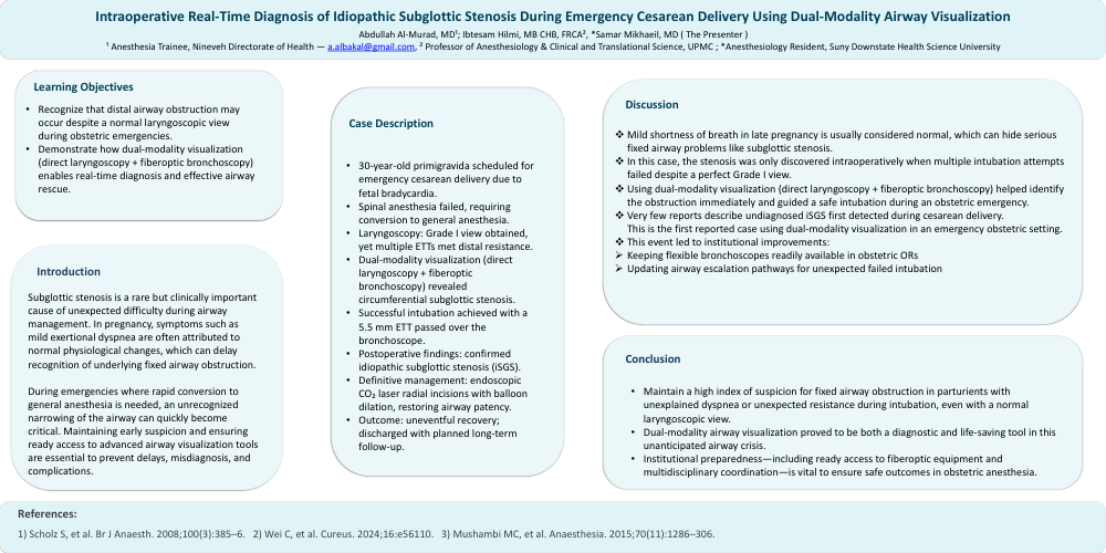
Intraoperative Real-Time Diagnosis of Idiopathic Subglottic Stenosis During Emergency Cesarean Delivery Using Dual-Modality Airway Visualization
Poster Presenter
Part of Topic
Airway - Obstetrics
Intraoperative Real-Time Diagnosis of Idiopathic Subglottic Stenosis During Emergency Cesarean Delivery Using Dual-Modality Airway Visualization
Abdullah Al-MuradMD1, Ibtesam Hilmi MB CHB, FRCA2
1 Anesthesia Department Trainee, Nineveh Directorate of Health; a.albakal@gmail.com
2 Professor of Anesthesiology and Professor of Clinical and Translational Science Institute, University of Pittsburgh Medical Center; hilmiia@anes.upmc.edu
Learning Objective
- Recognize that distal airway obstruction may present despite a normal laryngoscopic view during obstetric emergencies.
- Illustrate how combining direct laryngoscopy with fiberoptic bronchoscopy can facilitate real-time diagnosis and successful airway rescue.
Case Description
A 30-year-old primigravida required emergency cesarean delivery for fetal bradycardia. Spinal anesthesia failed, necessitating conversion to general anesthesia. Although laryngoscopy revealed a Grade I view, multiple endotracheal tubes could not be advanced due to distal resistance. Escalation to dual-modality visualization—integrating direct laryngoscopy with fiberoptic bronchoscopy—identified circumferential subglottic stenosis. A 5.5 mm endotracheal tube was successfully passed over the bronchoscope. Postoperative evaluation confirmed idiopathic subglottic stenosis, and the patient later underwent endoscopic CO₂ laser radial incisions with balloon dilation, restoring airway patency. She was discharged without complications, with long-term follow-up arranged.
This case demonstrates the critical importance of considering distal airway pathology when intubation fails despite a favorable laryngeal view. The combined use of direct and fiberoptic visualization enabled real-time diagnosis and safe airway management in a time-sensitive obstetric emergency.
Discussion
Mild exertional dyspnea during late pregnancy is often attributed to physiological changes, which can delay recognition of fixed airway lesions such as subglottic stenosis. In this case, idiopathic subglottic stenosis (iSGS) was only revealed intraoperatively when multiple intubation attempts failed despite a Grade I laryngoscopic view. Escalation to dual-modality assessment—direct laryngoscopy combined with fiberoptic bronchoscopy, enabled real-time diagnosis and safe airway rescue during a high-stakes obstetric emergency.
To our knowledge, very few reports describe intraoperative discovery of undiagnosed iSGS during cesarean delivery, and this appears to be the first case utilizing dual-modality visualization in an emergent obstetric setting. The event prompted institutional changes, including stocking flexible bronchoscopes in all obstetric ORs and revising airway escalation protocols. Clinicians must remain alert to the possibility of distal airway obstruction when intubation fails despite a normal glottic view and be prepared to escalate promptly, with early otolaryngology involvement ensuring definitive management and future peripartum planning.
References
- Scholz S, et al. Br J Anaesth. 2008;100(3):385–6.
- Wei C, et al. Cureus. 2024;16:e56110.
- Mushambi MC, et al. Anaesthesia. 2015;70(11):1286–306.