197 posters, 15 videos, 31 sessions, 510 authors
ePostersLive by SciGen Technologies S.A. All rights reserved.
2026 AO Annual Meeting
Conference ePosters

P208
Orthodontic, Implant and Regenerative Approach to a Mandibular Anterior Defect: 5-year Outcome
Poster Presenter
Authors
Part of Topic
Orthodontic, Implant, and Regenerative Approach to a Mandibular Anterior Defect: 5-Year Outcome
Jia Yuan, BDS, MS¹; Zhihui Mai, BDS, MS, PhD¹; Hom-Lay Wang, DDS, MSD, PhD²*
¹ Department of Stomatology, The Third Affiliated Hospital of Sun Yat-sen University, Guangzhou, China
² Department of Periodontics and Oral Medicine, University of Michigan School of Dentistry, Ann Arbor, MI, USA
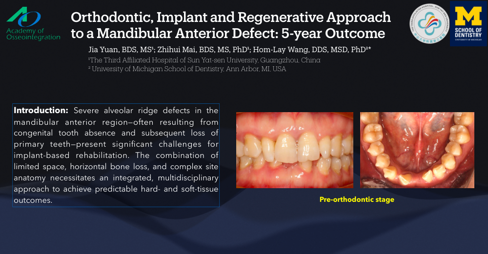
Background: Severe alveolar ridge defects in the mandibular anterior region—often resulting from congenital tooth absence and subsequent loss of primary teeth—present significant challenges for implant-based rehabilitation. The combination of limited space, horizontal bone loss, and complex site anatomy necessitates an integrated, multidisciplinary approach to achieve predictable hard- and soft-tissue outcomes.
Methods: A 30-year-old female presented with significant spacing after exfoliation of a retained primary mandibular anterior tooth, with missing #24 and #26, distal rotation of #25, and a deep overbite/overjet. Following 15 months of orthodontic space creation, severe horizontal ridge deficiency (approximately 6mm) was confirmed clinically and via CBCT. A single-stage regenerative protocol was performed: simultaneous guided bone regeneration (GBR) at implant sites (#23, #25) with bone level tapered implants and guided tissue regeneration (GTR) at adjacent natural teeth (#24, #26), utilizing deproteinized bovine bone mineral (DBBM)) xenograft and an absorbable collagen membrane for multi-wall defect regeneration. Tension-free primary closure was achieved.
Results: Healing was uneventful. Over five years, radiographic and clinical assessments confirmed stable bucco-lingual hard-tissue augmentation, increased keratinized mucosa, and healthy peri-implant soft-tissue contours. CBCT demonstrated robust lingual and buccal bone regeneration and thickness at both implant sites and natural teeth, with no mucosal or gingival recession and maintained bone volume.
Conclusion: This case illustrates that a coordinated approach combining orthodontic space creation with simultaneous GBR at implant sites and GTR at neighboring teeth, utilizing DBBM and collagen membrane, facilitates predictable, long-term rehabilitation of complex mandibular anterior defects. This strategy achieved symmetrical, three-dimensional bone augmentation and maintained stable functional and esthetic results over a five-year follow-up.