197 posters, 15 videos, 31 sessions, 510 authors
ePostersLive by SciGen Technologies S.A. All rights reserved.
2026 AO Annual Meeting
Conference ePosters

P114
Utilizing Post-operative CBCT to Enhance Pre-operative Planning Integration in Full-arch Implant Rehabilitation
Poster Presenter
Authors
Part of Topic
Utilizing Post-Operative CBCT to Enhance Pre-Operative Planning Integration in Full-Arch Implant Rehabilitation
INTRODUCTION
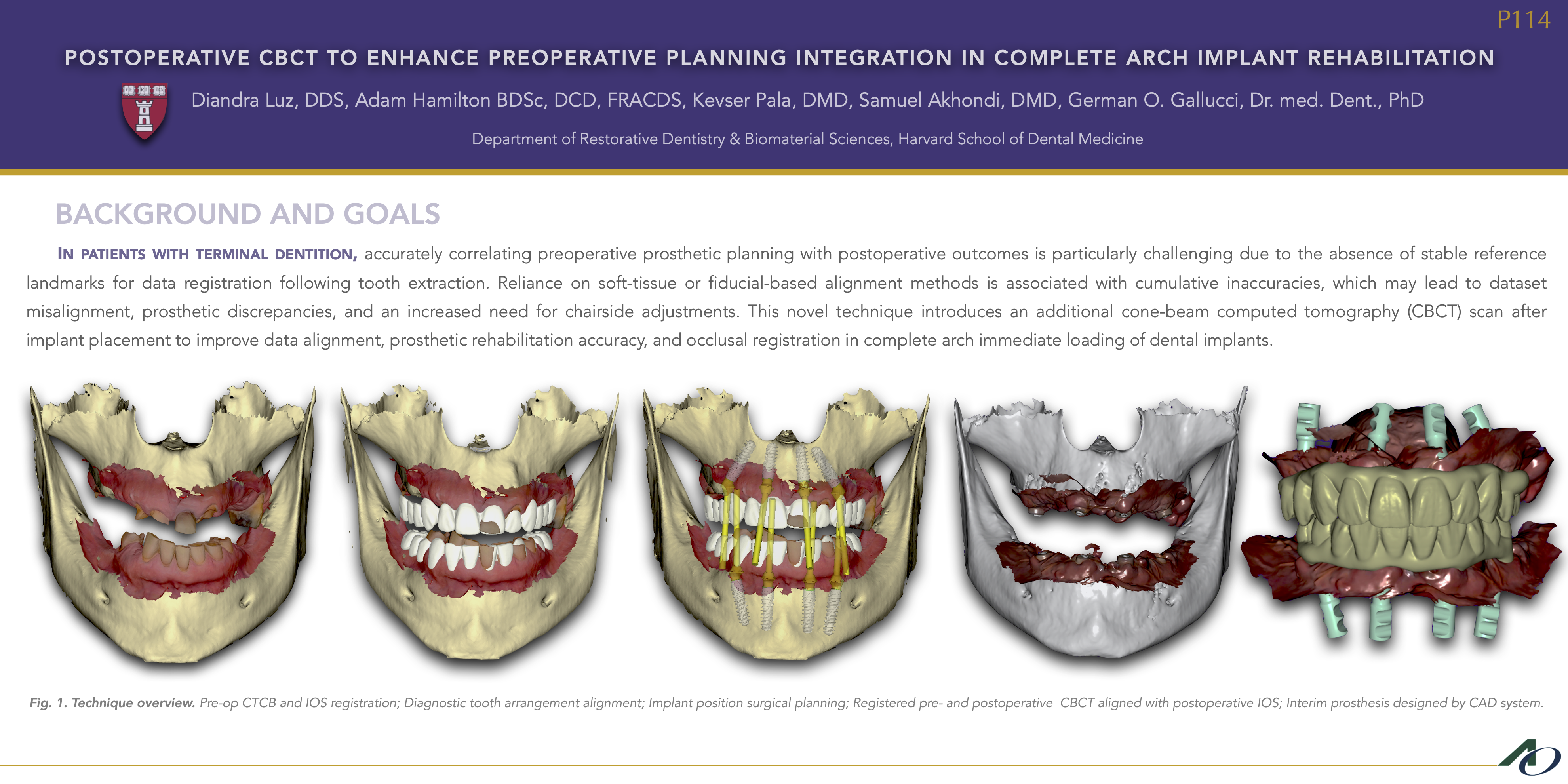
Rehabilitating terminal dentition with an immediately loaded, fixed implant-supported full-arch restoration offers a predictable solution and high patient satisfaction. However, after tooth extraction, vertical dimension information is lost, and reference structures for aligning initial prosthetic design with the post-extraction clinical situation are lacking. This complicates precise postoperative occlusal registration and ideal tooth positioning. The present workflow introduces pre-surgical and post-surgical CBCTs to improve data alignment, prosthetic rehabilitation, and occlusal registration for complete arch immediate loading of dental implants.
METHODS
Pre-surgical steps: 1) Digital impression (IOS) of the terminal dentition 2) Ideal digital tooth set-up design on CAD software 3) Pre-operative CBCT 4) Registration of pre-op IOS, CBCT and digital set-up (DATASET A) on CoDiagnostix 5) Digital planning of implant positioning 6) Implant placement 7) Immediate postoperative CBCT. Immediate provisionalization: 1) Post-op IOS to capture healing cap positions 2) Registration of post-op CBCT to post-op IOS (DATASET B) using the healing caps as reference 3) Export DATASET B 4) Registration of datasets A and B using bony references from CBCTs (i.e. inferior border of the mandible and zygomatic process of the maxilla) 5) DATASET B is digitally shifted into the correct occlusion using the pre-op set-up 6) Photogrammetry to ensure accuracy of implant placement 7) Adjustment of the provisional prosthesis to the actual implant position 8) Milling of provisional prostheses - PMMA 9) Delivery of provisional prostheses within 24 hours, with confirmed passive fit and occlusion.
RESULTS
The presented technique effectively transfers preoperative planning to the postoperative intraoral environment by using pre- and post-op CBCT scans with stable bony landmarks. No occlusal adjustments for mandibular and maxillary immediate prostheses were necessary using this approach.
CONCLUSION
While this technique requires an additional post-implant CBCT scan, it offers precise radiographic confirmation of implant positioning, ensuring accurate prosthetic alignment. Unlike removable appliances and fiduciary markers, prone to misalignment, this method allows mandibular and maxillary prostheses insertion with minimal occlusal adjustments and adapts to various workflows, including freehand implant placement.