226 posters, 5 topics, 20 sessions, 599 authors, 292 institutions
ePostersLive by SciGen Technologies S.A. All rights reserved.
Obstetric Anaesthesia Annual Scientific Meeting 2026
14-15 May 2026 | Liverpool Convention Centre

7162763
A diagnostic challenge: renal angle pain at 35 weeks gestation
Poster Presenter
Authors
Affiliations
Part of Topic
Case report
Introduction
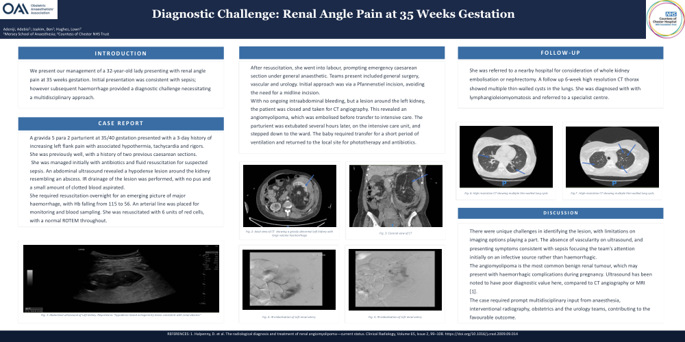
We present our management of a 32-year-old lady presenting with renal angle pain at 35 weeks gestation. Initial presentation was consistent with sepsis; however subsequent haemorrhage provided a diagnostic challenge necessitating a multidisciplinary approach.
Case Report
A gravida 5 para 2 parturient at 35/40 gestation presented with a 3-day history of increasing left flank pain with associated hypothermia, tachycardia and rigors. She was previously well, with a history of two previous caesarean sections.
She was managed initially with antibiotics and fluid resuscitation for suspected sepsis. An abdominal ultrasound revealed a hypodense lesion around the kidney resembling an abscess. IR drainage of the lesion was performed, with no pus and a small amount of clotted blood aspirated.
She required resuscitation overnight for an emerging picture of major haemorrhage, with Hb falling from 115 to 56. An arterial line was placed for monitoring and blood sampling. She was resuscitated with 6 units of red cells, with a normal ROTEM throughout.
After resuscitation, she went into labour, prompting emergency caesarean section under general anaesthetic. Teams present included general surgery, vascular and urology. Initial approach was via a Pfannenstiel incision, avoiding the need for a midline incision.
With no ongoing intraabdominal bleeding, but a lesion around the left kidney, the patient was closed and taken for CT angiography. This revealed an angiomyolipoma, which was embolised before transfer to intensive care. The parturient was extubated several hours later, on the intensive care unit, and stepped down to the ward. The baby required transfer for a short period of ventilation and returned to the local site for phototherapy and antibiotics.
Follow-Up
She was referred to a nearby hospital for consideration of whole kidney embolisationor nephrectomy. A follow up 6-week high resolution CT thorax showed multiple thin-walled cysts in the lungs. She was diagnosed with with lymphangioleiomyomatosis and referred to a specialist centre.
Discussion
There were unique challenges in identifying the lesion, with limitations on imaging options playing a part. The absence of vascularity on ultrasound, and presenting symptoms consistent with sepsis focusing the team’s attention initially on an infective source rather than haemorrhagic.
The angiomyolipoma is the most common benign renal tumour, which may present with haemorrhagic complications during pregnancy. Ultrasound has been noted to have poor diagnostic value here, compared to CT angiography or MRI [1].
The case required prompt multidisciplinary input from anaesthesia, interventional radiography, obstetrics and the urology teams, contributing to the favourable outcome.
References
1. Halpenny, D. et al. The radiological diagnosis and treatment of renal angiomyolipoma—current status. Clinical Radiology, Volume 65, Issue 2, 99–108. https://doi.org/10.1016/j.crad.2009.09.014