215 posters, 5 topics, 22 sessions, 596 authors, 283 institutions
ePostersLive by SciGen Technologies S.A. All rights reserved.
Obstetric Anaesthesia Annual Scientific Meeting 2025
May 15 - 16, 2025 | ICC, 2 Lanyon Place, Belfast BT1 3WH

6779745
Typical L2L3 and L3L4 interspinous space dimensions: Details that help guide epidural insertion.
Part of Topic
Original Research
Typical L2L3 and L3L4 Interspinous Space Dimensions: Details That Help Guide Epidural Insertion
Sue Lawrence1,2,3, Nicole Atcheson1, David Reutens1,2,3, David Sturgess2,4
Introduction
Midline translaminar insertion of a lumbar spinal or epidural is a simple procedure until it is not.[1] This morphometric summary assists to inform needle advancement, redirections and sonogram interpretation.
Methods
Ethics approval was obtained for examination and magnetic resonance imaging (MRI) of the lumbar spines of healthy young adults (18 – 20 years). MRI measurements were taken from a midline, mid-interspinous space axis for each interspace
(T12L1-L5S1). Values are given as mean and standard deviation (m, sd).
Results
The 27 volunteers (16 female, 11 male) were found to have normal lumbar anatomy.
L3L4 was the modal and highest level of the intercristal line (66.7%, n = 18).
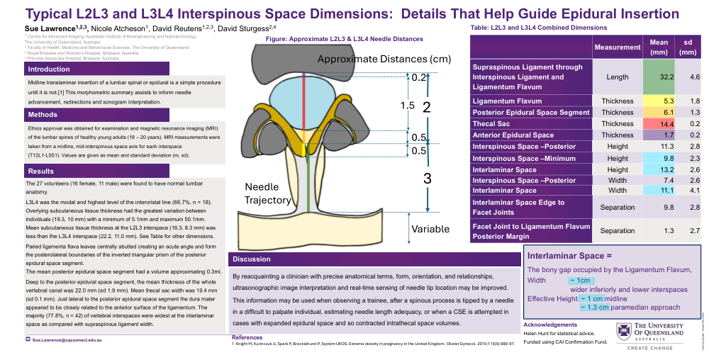
Overlying subcutaneous tissue thickness had the greatest variation between individuals (19.3, 10 mm) with a minimum of 5.1mm and maximum 50.1mm.
Mean subcutaneous tissue thickness at the L2L3 interspace (16.3, 8.3 mm) was less than the L3L4 interspace (22.2, 11.0 mm). See Table for other dimensions.
Paired ligamenta flava leaves centrally abutted creating an acute angle and form the posterolateral boundaries of the inverted triangular prism of the posterior epidural space segment.
The mean posterior epidural space segment had a volume approximating 0.3ml.
Deep to the posterior epidural space segment, the mean thickness of the whole vertebral canal was 22.0 mm (sd 1.9 mm). Mean thecal sac width was 19.4 mm (sd 0.1 mm). Just lateral to the posterior epidural space segment the dura mater appeared to be closely related to the anterior surface of the ligamentum. The majority (77.8%, n = 42) of vertebral interspaces were widest at the interlaminar space as compared with supraspinous ligament width.
Discussion
By reacquainting a clinician with precise anatomical terms, form, orientation, and relationships, ultrasonographic image interpretation and real-time sensing of needle tip location may be improved.
This information may be used when observing a trainee, after a spinous process is tipped by a needle in a difficult to palpate individual, estimating needle length adequacy, or when a CSE is attempted in cases with expanded epidural space and so contracted intrathecal space volumes.
Interlaminar Space =
The bony gap occupied by the Ligamentum Flavum,
Width ~ 1cm
wider inferiorly and lower interspaces
Effective Height ~ 1 cm midline
~ 1.3 cm paramedian approach