293 posters, 56 videos, 24 topics, 13 sessions, 658 authors, 215 institutions
ePostersLive by SciGen Technologies S.A. All rights reserved.
ISMICS 2025 Annual Meeting
28 - 31 May, 2025 | Istanbul, Turkey

D175
Robot-Assisted Ivor Lewis Esophagectomy: A Case Report
Poster Presenter
Part of Topic
Thoracic Surgery – Esophageal
Robot-Assisted Ivor Lewis Esophagectomy: A Case Report
Serkan UYSAL¹, Ilknur AYDAR¹, Mehmet Ruchan KARACA², Ulas KUMBASAR¹, Nezih AKKAPULU²
¹Department of Thoracic Surgery, Hacettepe University, Ankara, Türkiye
²Department of General Surgery, Hacettepe University, Ankara, Türkiye
BACKGROUND
Esophageal cancer ranks among the top ten cancers worldwide and is associated with poor prognosis and high mortality. Most patients present with progressive dysphagia, weight loss, and nutritional deterioration, often leading to delayed diagnosis. Surgical resection remains the cornerstone of curative treatment in operable cases. Among the available techniques, Ivor Lewis esophagectomy, which involves a two-phase resection via abdominal and thoracic approaches, is widely used for tumors of the middle and distal esophagus. However, the open version of this procedure is associated with considerable postoperative pain, pulmonary complications, and prolonged recovery. In recent years, robot-assisted esophagectomy has emerged as a promising minimally invasive alternative, offering enhanced precision, better visualization, and reduced trauma to surrounding tissues. Studies have shown that robotic approaches are associated with reduced blood loss, shorter hospital stays, and lower postoperative morbidity compared to open surgery. In this report, we present a case of totally robotic Ivor Lewis esophagectomy, highlighting its feasibility, technical considerations, and potential benefits for patients with resectable esophageal cancer.
METHODS
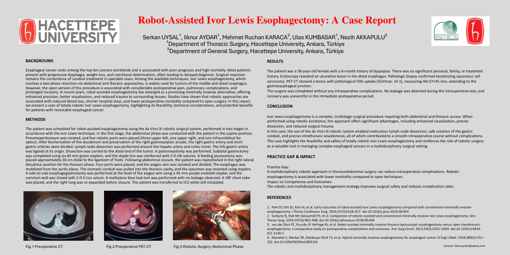
The patient was scheduled for robot-assisted esophagectomy using the da Vinci Xi robotic surgical system, performed in two stages in accordance with the Ivor Lewis technique. In the first stage, the abdominal phase was conducted with the patient in the supine position. Pneumoperitoneum was created, and five robotic ports were placed (three upper left, one upper right, and one infraumbilical for optics). After Kocherization of the duodenum and preservation of the right gastroepiploic arcade, the right gastric artery and short gastric arteries were divided. Lymph node dissection was performed around the hepatic artery and celiac trunk. The left gastric artery was ligated at its origin. Dissection was carried to the distal end of the tumor. A pyloromyotomy was performed. Subtotal gastrectomy was completed using six 60 mm green staplers, and the staple line was reinforced with 2-0 silk sutures. A feeding jejunostomy was placed approximately 30 cm distal to the ligament of Treitz. Following abdominal closure, the patient was repositioned in the right lateral decubitus position for the thoracic phase. Four ports were placed, and the azygos vein was isolated and divided. The esophagus was mobilized from the aortic plane. The stomach conduit was pulled into the thoracic cavity, and the specimen was resected using staplers. A side-to-side esophagogastrostomy was performed at the level of the azygos vein using a 45 mm purple endoGIA stapler, and the common wall was closed with 2-0 V-Loc suture. A methylene blue leak test was performed with no leakage observed. A 28F chest tube was placed, and the right lung was re-expanded before closure. The patient was transferred to ICU while still intubated.
RESULTS
The patient was a 58-year-old female with a 6-month history of dyspepsia. There was no significant personal, family, or treatment history. Endoscopy revealed an ulcerative lesion in the distal esophagus. Pathologic biopsy confirmed keratinizing squamous cell carcinoma. PET-CT showed a lesion with pathological FDG uptake (SUVmax: 14.1), measuring 40×27×45 mm, extending to the gastroesophageal junction. The surgery was completed without any intraoperative complications. No leakage was detected during the intraoperative test, and recovery was uneventful in the immediate postoperative period.
CONCLUSION
Ivor Lewis esophagectomy is a complex, multistage surgical procedure requiring both abdominal and thoracic access. When performed using robotic assistance, this approach offers significant advantages, including enhanced visualization, precise dissection, and reduced surgical trauma. In this case, the use of the da Vinci Xi robotic system enabled meticulous lymph node dissection, safe creation of the gastric conduit, and precise intrathoracic anastomosis, all of which contributed to a smooth intraoperative course without complications. This case highlights the feasibility and safety of totally robotic Ivor Lewis esophagectomy and reinforces the role of robotic surgery as a valuable tool in managing complex esophageal cancers in a multidisciplinary surgical setting.