293 posters, 56 videos, 24 topics, 13 sessions, 658 authors, 215 institutions
ePostersLive by SciGen Technologies S.A. All rights reserved.
ISMICS 2025 Annual Meeting
28 - 31 May, 2025 | Istanbul, Turkey

P99
Robotic Triple Valve Surgery: Operative Setup And Technique
Part of Topic
Video
Background:
Robotic mitral valve surgery is well-defined, robotic multiple valve surgery is rare and robotic triple valve surgery is a new concept. Robotic aortic valve replacement, mitral valve repair, tricuspid valve repair, patent foramen ovale and atrial appendage closure were performed in our center to a 65 year-old female with a history of hypertension, atrial fibrillation and diabetes mellitus. She had a BMI of 38.6 kg/m2. Echocardiography revealed severe mitral regurgitation with annular dilatation, severe tricuspid regurgitation, severe aortic stenosis with calcified aortic leaflets, and a small aortic annulus (21 mm) with an EF of 40%. The left atrium was severely enlarged.
Operative Setup and Technique:
TEE-guided cannulation of right axillary artery, right femoral vein and right jugular vein was performed. Severe aortic valve stenosis, severe mitral valve and tricuspid valve regurgitation were confirmed with TEE.
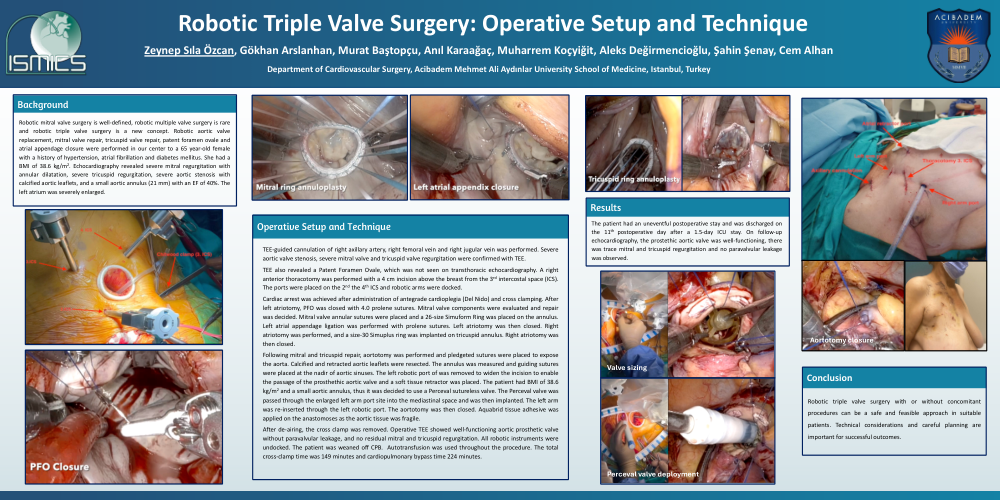
TEE also revealed a Patent Foramen Ovale, which was not seen on transthoracic echocardiography. A right anterior thoracotomy was performed with a 4 cm incision above the breast from the 3rd intercostal space (ICS). The ports were placed on the 2nd the 4th ICS and robotic arms were docked.
Cardiac arrest was achieved after administration of antegrade cardioplegia (Del Nido) and cross clamping. After left atriotomy, PFO was closed with 4.0 prolene sutures. Mitral valve components were evaluated and repair was decided. Mitral valve annular sutures were placed and a 26-size Simuform Ring was placed on the annulus. Left atrial appendage ligation was performed with prolene sutures. Left atriotomy was then closed. Right atriotomy was performed, and a size-30 Simuplus ring was implanted on tricuspid annulus. Right atriotomy was then closed.
Following mitral and tricuspid repair, aortotomy was performed and pledgeted sutures were placed to expose the aorta. Calcified and retracted aortic leaflets were resected. The annulus was measured and guiding sutures were placed at the nadir of aortic sinuses. The left robotic port of was removed to widen the incision to enable the passage of the prosthethic aortic valve and a soft tissue retractor was placed. The patient had BMI of 38.6 kg/m2 and a small aortic annulus, thus it was decided to use a Perceval sutureless valve. The Perceval valve was passed through the enlarged left arm port site into the mediastinal space and was then implanted. The left arm was re-inserted through the left robotic port. The aortotomy was then closed. Aquabrid tissue adhesive was applied on the anastomoses as the aortic tissue was fragile.
After de-airing, the cross clamp was removed. Operative TEE showed well-functioning aortic prosthetic valve without paravalvular leakage, and no residual mitral and tricuspid regurgitation. All robotic instruments were undocked. The patient was weaned off CPB. Autotransfusion was used throughout the procedure. The total cross-clamp time was 149 minutes and cardiopulmonary bypass time 224 minutes.
Results:
The patient had an uneventful postoperative stay and was discharged on the 11th postoperative day after a 1.5-day ICU stay. On follow-up echocardiography, the prostethic aortic valve was well-functioning, there was trace mitral and tricuspid regurgitation and no paravalvular leakage was observed.
Conclusion:
Robotic triple valve surgery with or without concomitant procedures can be a safe and feasible approach in suitable patients. Technical considerations and careful planning are important for successful outcomes.