225 posters, 34 videos, 18 topics, 14 sessions, 251 authors, 55 institutions
ePostersLive by SciGen Technologies S.A. All rights reserved.
ISMICS 2024 Annual Meeting
29 May - 1 June, 2024 | Athens, Greece

D.50
Three Dimensional Virtual Reality Evaluation In Cardiovascular Surgery
Poster Presenter
Authors
Part of Topic
Education, Imaging and Facilitating Technologies
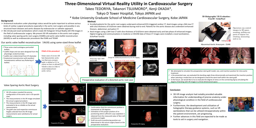
Three-Dimensional Virtual Reality Utility in Cardiovascular Surgery
Background:
ØAnatomical evaluation under physiologic status would be quite important to achieve various kinds of cardiac surgical procedures especially in the aortic root surgery and possibly in any structural heart diseases and aortic diseases by endovascular or catheter approach.
ØWe introduced novel workstations which create 3D Hologram Virtual Reality (3D-VR) Images in the field of cardiovascular surgery. We present 3D-VR evaluation in the aortic root surgery including valve sparing aortic root surgery (VSARS) and aortic valve leaflet reconstruction (AVLRS) as well as endovascular procedures like EVAR and TEVAR
Method:
ØEnrolled patients for the aortic root surgery underwent enhanced ECG-triggered cardiac CT. Axial images using a 264-row CT with slice thickness of 0.625mm were obtained during mid-to-end. Patients for the aortic disease also underwent enhanced thoracic, abdominal, and pelvic CT.
ØAxial images using a 264-row CT with slice thickness of 0.625mm were obtained early and late phase of enhanced images. Digital imaging and communications in medicine or DICOM data of these CT images were installed a novel workstation, EchoPixel Ture3D.
Our aortic valve leaflet reconstruction(AVLR) using same-sized three leaflet
üThree same-sized autologous pericardial leaflets
üLeaflet shape and size were designed from the physiologic anatomical data
üLeaflet size is determined from STJ diameter
üAnatomical reconstruction with excellent hemodynamics without any fluttering of leaflets
Abcominal Aortic Aneurysm : EVAR
üWe attempted to simulate the preoperative stent graft model, size, and insertion position for intra-aortic treatment.
ün the presented case, we evaluated the bending angle three-dimensionally and examined the insertion position when selecting a model that can be designed to bend the stent itself within the stent graft.
üIn the future, we would like to try to evaluate the degree of stress on the connecting leg by simulating the degree of bending with VR images in a three-dimensional space.
Valve Sparing Aortic Root Surgery
ü3D-VR analysis revealed the precise configuration of the aortic root in 3D spacidic view
üThe measurements in the VR image simulated the actual surgical procedure.
üThe measurements in the VR image were evaluated to be as reliable as the actual measurements.
üThe graft sizes and respective commissure heights selected from the VR image measurements were satisfactory for actual surgery.
Conclusion
Ø3D-VR image analysis had notably provided valuable information for understanding of precise anatomy under physiological condition in the field of cardiovascular surgery.
ØFurthermore, the development and utilization of holographic therapy guidance systems, such as VR workstations that can be utilized intraoperatively within the patient environment, are progressing.
ØFurther advances in this field are expected to be made as tools to aid in surgery and navigation.