225 posters, 34 videos, 18 topics, 14 sessions, 251 authors, 55 institutions
ePostersLive by SciGen Technologies S.A. All rights reserved.
ISMICS 2024 Annual Meeting
29 May - 1 June, 2024 | Athens, Greece
PC.122
RIGHT VIDEO-ASSISTED THORACOSCOPIC SURGICAL APPROACH TO A GIANT SOLITARY FIBROUS TUMOR OF THE ESOPHAGUS
Poster Presenter
Authors
Part of Topic
Thoracic Surgery
Video
RIGHT VIDEO-ASSISTED THORACOSCOPIC SURGICAL APPROACH TO A GIANT SOLITARY FIBROUS TUMOR OF THE ESOPHAGUS
Aroub Alkaaki MD FRCSC1, Elisabeth Savonitto, MD2, Sebastien Gilbert, MD FRCSC1
1 University of Ottawa, Ottawa, ON, Canada, 2 Dalhousie University, Halifax, Dalhousie University, NS, Canada
BACKGROUND
Solitary fibrous tumours (SFTs) are fibroblastic mesenchymal neoplasms commonly found in the visceral pleura, upper respiratory tract, and lungs.1 However, their presence in the esophagus is exceptionally rare, with only a few reported cases. 2-4 This video outlines our specific surgical approach for treating this rare tumour.
METHODS
A case report supplemented by a video presentation was prepared after obtaining informed consent from the patient.
CASE SUMMARY
• 57-year-old man with a history of dysphagia to solid food for two years.
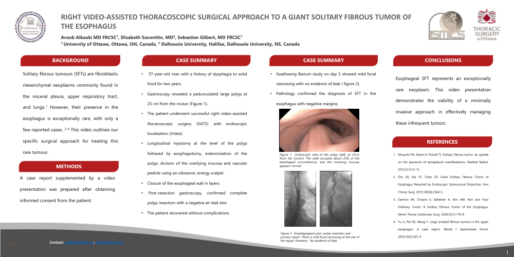
•Gastroscopy revealed a pedunculated large polyp at 25 cm from the incisor (Figure 1).
•The patient underwent successful right video-assisted thoracoscopic surgery (VATS) with endoscopic localization (Video).
•Longitudinal myotomy at the level of the polyp followed by esophagotomy, exteriorization of the polyp, division of the overlying mucosa and vascular pedicle using an ultrasonic energy scalpel.
•Closure of the esophageal wall in layers.
• Post-resection gastroscopy, confirmed complete polyp resection with a negative air leak test.
•The patient recovered without complications.
•Swallowing Barium study on day 5 showed mild focal narrowing with no evidence of leak ( figure 2).
•Pathology confirmed the diagnosis of SFT in the esophagus with negative margins.
CONCLUSION
Esophageal SFT represents an exceptionally rare neoplasm. This video presentation demonstrates the viability of a minimally invasive approach in effectively managing these infrequent tumors
REFERENCES