167 posters, 28 videos, 9 topics, 12 sessions, 337 authors, 160 institutions
ePostersLive by SciGen Technologies S.A. All rights reserved.
ISMICS 2023 Annual Meeting
31 May - 3 June, 2023 | Boston, MA

PC76
Utility Of Robot-assisted Bronchoscopy With Radial Endobronchial Ultrasound For Marking Of Subcentimeter Pulmonary Nodules For Resection
Poster Presenter
Authors
Anna Xue, Devon Anderson, Sarah Chen, Chinh Phan, Lisa Brown, David Cooke, Luis Godoy
Affiliations
Part of Topic
Thoracic Surgery
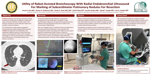
Centrally located subcentimeter pulmonary nodules suspicious for malignancy are difficult to localize and resect minimally invasively and often require more extensive resection to ensure adequate margins and complete resection of disease. The introduction of robot-assisted navigational bronchoscopy with radial endobronchial ultrasound (r-EBUS) has allowed for the identification and subsequent sublobar resection of these pulmonary nodules. We evaluated the utility of this innovative technology to mark and resect pulmonary nodules that would otherwise be unresectable without more extensive resections.
Preoperative dedicated high-definition computerized tomography (CT) was performed in all patients. Virtual bronchoscopic images were then generated using the planning software and lung registration points were marked on the virtual image intraoperatively. Next, the navigation phase is initiated to locate the target lesion and robot-assisted bronchoscopy is used to position the transbronchial needle. Fluoroscopy and r-EBUS are then used to guide the transbronchial needle to inject a mixture of indocyanine green and methylene blue to mark the target lesion and to create a track for coil or fiducial marker placement, allowing subsequent minimally invasive sublobar lung resection.
Robot-assisted bronchoscopy with r-EBUS localization of pulmonary nodules via delivery of dye agents, placement of coils, and/or fiducial markers have aided in multiple successful minimally invasive sublobar resection of subcentimeter and partially solid pulmonary nodules. Intraoperative visualization of the marked lesions allows for precise resection of the target lesions with appropriate margins on pathology.
This emerging technology has enabled successful minimally invasive lung sparing sublobar resection with appropriate margins for partially solid subcentimeter pulmonary nodules in all segments of the lung that traditionally are difficult to locate.