477 posters, 14 topics, 2,065 authors, 1,055 institutions
ePostersLive by SciGen Technologies S.A. All rights reserved.
The Joint EBJIS-MSIS 2026 Conference
17 - 19 September, 2026 | Porto, Portugal

EP339
Where in the peri-prosthetic membrane of PJI is Staphylococcus aureus located? A digital pathology study
Poster Presenter
Authors
Sophie Amalie Blirup-Plum, Mats Bue, Christen Ravn, Louise Kruse Jensen
Affiliations
Department of Clinical Medicine, Aarhus University, Aarhus, Denmark; Department of Orthopaedic Surgery, Aarhus University Hospital, Aarhus, Denmark, University Of Copenhagen, Department of Veterinary and Animal Sciences, University Of Copenhagen, Frederiksberg, Denmark, Department of Orthopaedic Surgery, Aarhus University Hospital, Aarhus, Denmark
Part of Topic
Prosthetic Joint Infections
Where in the peri-prosthetic membrane of PJI is Staphylococcus aureus located?
A digital pathology study
BACKGROUND
Sampling the periprosthetic membrane for microbiological and histological analysis is recommended to diagnose periprosthetic joint infection (PJI). Microbiology identifies the pathogen by culture, whereas histology assesses neutrophil infiltration but rarely detects bacteria.
Aim: To use immunohistochemistry (IHC) and digital pathology to investigate the location of Staphylococcus aureus within the membrane.
STUDY DESIGN
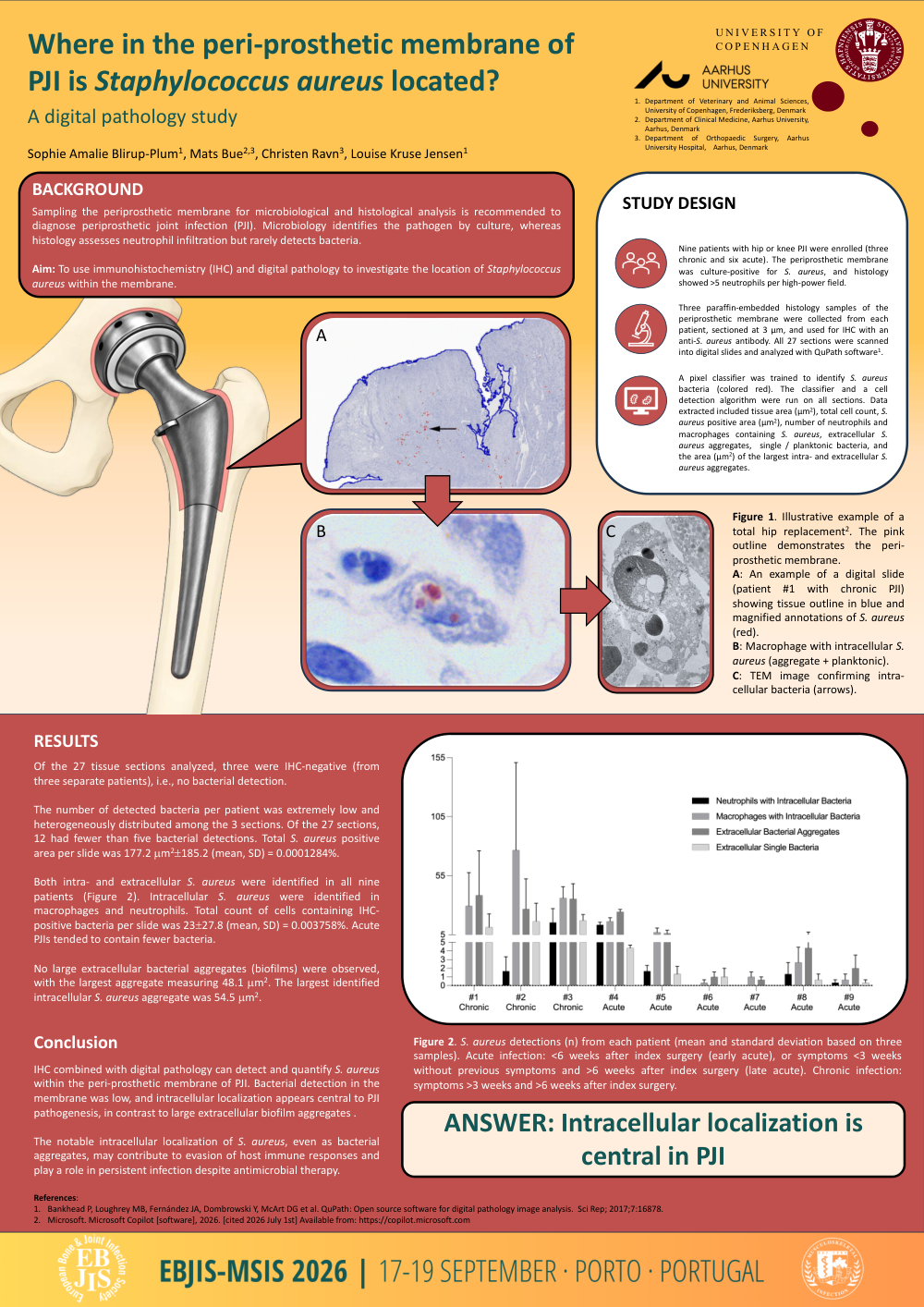
Nine patients with hip or knee PJI were enrolled (three chronic and six acute). The periprosthetic membrane was culture-positive for S. aureus, and histology showed >5 neutrophils per high-power field.
Three paraffin-embedded histology samples of the periprosthetic membrane were collected from each patient, sectioned at 3 µm, and used for IHC with an anti-S. aureus antibody. All 27 sections were scanned into digital slides and analyzed with QuPath software1.
A pixel classifier was trained to identify S. aureus bacteria (colored red). The classifier and a cell detection algorithm were run on all sections. Data extracted included tissue area (µm2), total cell count, S. aureus positive area (µm2), number of neutrophils and macrophages containing S. aureus, extracellular S. aureus aggregates, single / planktonic bacteria, and the area (µm2) of the largest intra- and extracellular S. aureus aggregates.
RESULTS
Of the 27 tissue sections analyzed, three were IHC-negative (from three separate patients), i.e., no bacterial detection.
The number of detected bacteria per patient was extremely low and heterogeneously distributed among the 3 sections. Of the 27 sections, 12 had fewer than five bacterial detections. Total S. aureus positive area per slide was 177.2 mm2±185.2 (mean, SD) = 0.0001284%.
Both intra- and extracellular S. aureus were identified in all nine patients (Figure 2). Intracellular S. aureus were identified in macrophages and neutrophils. Total count of cells containing IHC-positive bacteria per slide was 23±27.8 (mean, SD) = 0.003758%. Acute PJIs tended to contain fewer bacteria.
No large extracellular bacterial aggregates (biofilms) were observed, with the largest aggregate measuring 48.1 mm2. The largest identified intracellular S. aureus aggregate was 54.5 mm2.
CONCLUSION
IHC combined with digital pathology can detect and quantify S. aureus within the peri-prosthetic membrane of PJI. Bacterial detection in the membrane was low, and intracellular localization appears central to PJI pathogenesis, in contrast to large extracellular biofilm aggregates .
The notable intracellular localization of S. aureus, even as bacterial aggregates, may contribute to evasion of host immune responses and play a role in persistent infection despite antimicrobial therapy.
Figure 1.
Illustrative example of a total hip replacement2. The pink outline demonstrates the peri-prosthetic membrane. A: An example of a digital slide (patient #1 with chronic PJI) showing tissue outline in blue and magnified annotations of S. aureus (red). B: Macrophage with intracellular S. aureus (aggregate + planktonic). C: TEM image confirming intra-cellular bacteria (arrows).
Figure 2.
S. aureus detections (n) from each patient (mean and standard deviation based on three samples). Acute infection: <6 weeks after index surgery (early acute), or symptoms <3 weeks without previous symptoms and >6 weeks after index surgery (late acute). Chronic infection: symptoms >3 weeks and >6 weeks after index surgery.