477 posters, 14 topics, 2,052 authors, 1,056 institutions
ePostersLive by SciGen Technologies S.A. All rights reserved.
The Joint EBJIS-MSIS 2026 Conference
17 - 19 September, 2026 | Porto, Portugal

EP309
Deep Infection as a Major Complication of 3D-Printed Custom Prosthetic Reconstruction After Pelvic Bone Sarcoma Resection
Part of Topic
Prosthetic Joint Infections
BACKGROUND
Pelvic reconstruction after periacetabular tumor resection remains one of the most demanding procedures in orthopedic oncology. Custom 3D-printed implants improve anatomical matching and fixation, but the biological challenge is unchanged: extensive soft-tissue dissection, large dead space, long operating times and a large prosthetic surface all increase the risk of wound failure and deep infection [1–3]. A recent meta-analysis of 2,199 PI–PIII pelvic resections reported pooled rates of 15% for infection, 13% for wound-healing problems and 14% for implant revision or removal [2].
AIM
To describe the full course of wound complications and deep infection after custom 3D-printed hemipelvic reconstruction, rather than limiting the analysis to the first postoperative event. The primary focus was infection-related implant loss; microbiological findings were reviewed descriptively because the number of septic failures was too small for organism-specific statistical analysis.
PATIENTS AND METHODS
• Retrospective single-center series of 24 consecutive custom hemipelvic reconstructions performed between 21 August 2013 and 15 July 2024.
• Twenty-two patients had a malignant pelvic bone sarcoma and two had an aggressive/benign primary bone tumor. All reconstructions involved the periacetabular region and used patient-specific cutting guides and custom 3D-printed titanium implants according to the previously published institutional workflow [1].
• Wound-related reoperation, deep infection, debridement, implant removal and the reason for explanations were reviewed longitudinally. Microbiological cultures were considered as part of the infection assessment and interpreted descriptively.
• Because of the small number of septic events, no pathogen-specific risk analysis was attempted.
RESULTS
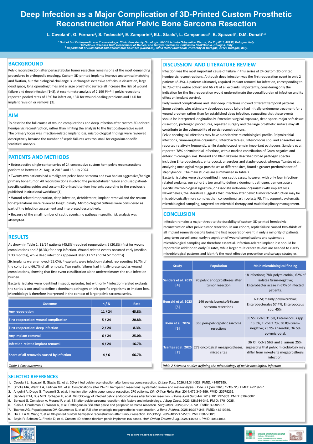
As shown in Table 1, 11/24 patients (45.8%) required reoperation: 5 (20.8%) first for wound complications and 2 (8.3%) for deep infection. Wound-related events occurred early (median 1.33 months), while deep infections appeared later (12.57 and 34.57 months).
Six implants were removed (25.0%); 4 explants were infection-related, representing 16.7% of the cohort and 66.7% of all removals. Two septic failures had initially presented as wound complications, showing that first-event classification alone underestimates the true infection burden.
Bacterial isolates were identified in septic episodes, but with only 4 infection-related explants the series is too small to define a dominant pathogen or link specific organisms to implant loss. Microbiology is therefore interpreted in the context of larger pelvic sarcoma series.
DISCUSSION AND LITERATURE REVIEW
Infection was the most important cause of failure in this series of 24 custom 3D-printed hemipelvicreconstructions. Although deep infection was the first reoperation event in only 2 patients (8.3%), 4 patients ultimately required implant removal for infection, corresponding to 16.7% of the entire cohort and 66.7% of all explants. Importantly, considering only the indication for the first reoperation would underestimate the overall burden of infection and its effect on implant survival.
Early wound complications and later deep infections showed different temporal patterns. Some patients who ultimately developed septic failure had initially undergone treatment for a wound problem rather than for established deep infection, suggesting that these events should be interpreted longitudinally. Extensive surgical exposure, dead space, major soft-tissue dissection, prolonged procedures, repeated surgery and the large prosthetic surface may all contribute to the vulnerability of pelvic reconstructions.
Pelvic oncological infections may have a distinctive microbiological profile. Polymicrobial infections, Gram-negative organisms, Enterobacterales, Enterococcus spp. and anaerobes are reported relatively frequently, while staphylococci remain important pathogens. Sanders et al. reported 78% polymicrobial infections, with a marked contribution of Gram-negative and enteric microorganisms. Bensaid and Klein likewise described broad pathogen spectra including Enterobacterales, enterococci, anaerobes and staphylococci, whereas Tsantes et al., analyzingoncological mega-prostheses at different sites, found a greater predominance of staphylococci. The main studies are summarized in Table 2.
Bacterial isolates were also identified in our septic cases; however, with only four infection-related explants, our cohort is too small to define a dominant pathogen, demonstrate a specific microbiological signature, or associate individual organisms with implant loss. Nevertheless, the literature suggests that infection after pelvic tumorreconstruction may be microbiologically more complex than conventional arthroplasty PJI. This supports systematic microbiological sampling, targeted antimicrobial therapy and multidisciplinary management.
CONCLUSION
Infection remains a major threat to the durability of custom 3D-printed hemipelvic reconstruction after pelvic tumor resection. In our cohort, septic failure caused two-thirds of all implant removals despite being the first reoperation event in only a minority of patients. Long-term surveillance, early recognition of wound complications and systematic microbiological sampling are therefore essential. Infection-related implant loss should be reported in addition to early PJI rates, while larger multicenterstudies are needed to clarify microbiological patterns and identify the most effective prevention and salvage strategies.
SELECTED REFERENCES
1.Cevolani L, Spazzoli B, Staals EL, et al. 3D-printed pelvic reconstruction after bone sarcoma resection. Orthop Surg. 2026;18:311-321. PMID: 41457850.
2.Smolle MA, Wenzl FA, Laitinen MK, et al. Complications after PI–PIII hemipelvic resections: systematic review and meta-analysis. Bone JtOpen. 2026;7:713-723. PMID: 42219227.
3.Angelini A, Drago G, Trovarelli G, et al. Infection after pelvic bone tumour resection: 270 patients. Clin Orthop Relat Res. 2014;472:349-359. PMID: 23975252.
4.Sanders PTJ, Bus MPA, Scheper H, et al. Microbiology of infected pelvic endoprostheses after tumour resection. J Bone Joint Surg Am. 2019;101:797-803. PMID: 31045667.
5.Bensaid S, Contejean A, Morand P, et al. SSI after pelvic sarcoma resection: risk factors and microbiology. J Surg Oncol. 2023;128:344-349. PMID: 37010035.
6.Klein A, Chudamani C, Wieser A, et al. Pathogens in SSI after pelvic and peripelvic sarcoma resection. Surg Infect. 2024;25:737-741. PMID: 39292207.
7.Tsantes AG, Papadopoulos DV, Goumenos S, et al. PJI after oncologic megaprosthetic reconstruction. J Bone Jt Infect. 2025;10:337-345. PMID: 41210930.
8.Hu X, Lu M, Wang Y, et al. 3D-printed custom hemipelvic reconstruction after tumour resection. Int Orthop. 2024;48:2217-2231. PMID: 38775826.
9.Boyle R, Scholes C, Franks D, et al. Custom 3D-printed titanium pelvic implants: 106 cases. ArchOrthop Trauma Surg. 2025;145:431. PMID: 40874964.