477 posters, 14 topics, 2,065 authors, 1,055 institutions
ePostersLive by SciGen Technologies S.A. All rights reserved.
The Joint EBJIS-MSIS 2026 Conference
17 - 19 September, 2026 | Porto, Portugal

EP188
A novel antimicrobial Iodine-treated orthopaedic implant surface demonstrates biocompatibility and improved osseointegration in a sheep ingrowth model
Poster Presenter
Authors
Devendra Gorhe, Lucia Pontiroli, Xiaoou Fan, Michael Maynard, Imran Khan
Affiliations
Part of Topic
Miscellaneous
Background: Periprosthetic joint infection (PJI) remains one of the most challenging unmet needs in orthopaedics. A novel antimicrobial Iodine surface treatment, applied to Ti6Al4V implants, may support the management of PJI by reducing microbial attachment and biofilm formation on the implant surface through the gradual release of iodine into the surrounding joint space. With the addition of an antimicrobial to the surface of a device, its biological safety must not be compromised. This study evaluated the local tissue response, acute systemic toxicity, genotoxicity and osseointegration effects of this new technology.
Objectives: The goal of this study was to assess the biocompatibility of Iodine-treated implants following the principles of ISO 10993 standards. The sheep implantation study included also blood and urine iodine levels and thyroid function markers.
Materials and Methods:
Iodine treatment: All test samples received the Iodine surface treatment via an electrochemical process developed from the work of Takaya et al.1
Cytotoxicity, irritation and sensitization: Extracts were obtained using a pour-to-cover method from Iodine-treated hip stems. For cytotoxicity, cells were incubated at 37°C for 48h, and then graded based on cellular characteristics and % lysis. For irritation, extracts were delivered via intracutaneous injections and observations for erythema and edema were noted at 24, 48 and 72 hours after injection. For sensitization, extracts were delivered intradermally and as soaked patches, and animals were scored based on dermal reactions.
Acute systemic toxicity: Iodine-treated acetabular cups were extracted using a 3 cm2/mL ratio. The extracts were injected intravenously (saline) or intraperitoneally (sesame oil), and the animals were observed and weighed for 72h.
Genotoxicity: Iodine-treated acetabular cups were extracted using a 3 cm2/mL ratio. Both the Mouse Lymphoma Assay (MLA) and the Reverse Mutation Assay (Ames) were performed.
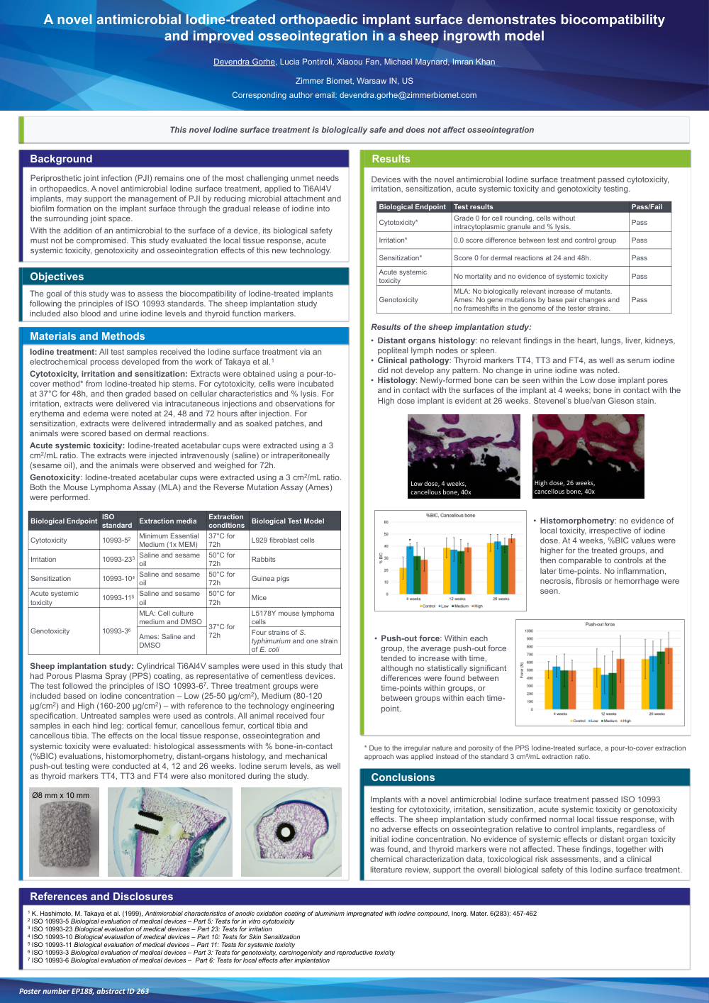
Sheep implantation study: Cylindrical Ti6Al4V samples were used in this study that had Porous Plasma Spray (PPS) coating, as representative of cementless devices. The test followed the principles of ISO 10993-6. Three treatment groups were included based on iodine concentration – Low (25-50 μg/cm2), Medium (80-120 μg/cm2) and High (160-200 μg/cm2) – with reference to the technology engineering specification. Untreated samples were used as controls. All animal received four samples in each hind leg: cortical femur, cancellous femur, cortical tibia and cancellous tibia. The effects on the local tissue response, osseointegration and systemic toxicity were evaluated: histological assessments with % bone-in-contact (%BIC) evaluations, histomorphometry, distant-organs histology, and mechanical push-out testing were conducted at 4, 12 and 26 weeks. Iodine serum levels, as well as thyroid markers TT4, TT3 and FT4 were also monitored during the study.
Results:
Devices with the novel antimicrobial Iodine surface treatment passed cytotoxicity, irritation, sensitization, acute systemic toxicity and genotoxicity testing.
Results of the sheep implantation study:
• Distant organs histology: no relevant findings in the heart, lungs, liver, kidneys, popliteal lymph nodes or spleen.
• Clinical pathology: Thyroid markers TT4, TT3 and FT4, as well as serum iodine did not develop any pattern. No change in urine iodine was noted.
• Histology: Newly-formed bone can be seen within the Low dose implant pores and in contact with the surfaces of the implant at 4 weeks; bone in contact with the High dose implant is evident at 26 weeks. Stevenel’s blue/van Gieson stain.
• Histomorphometry: no evidence of local toxicity, irrespective of iodine dose. At 4 weeks, %BIC values were higher for the treated groups, and then comparable to controls at the later time-points. No inflammation, necrosis, fibrosis or hemorrhage were seen.
• Push-out force: Within each group, the average push-out force tended to increase with time, although no statistically significant differences were found between time-points within groups, or between groups within each time-point.
Conclusions: Implants with a novel antimicrobial Iodine surface treatment passed ISO 10993 testing for cytotoxicity, irritation, sensitization, acute systemic toxicity or genotoxicity effects. The sheep implantation study confirmed normal local tissue response, with no adverse effects on osseointegration relative to control implants, regardless of initial iodine concentration. No evidence of systemic effects or distant organ toxicity was found, and thyroid markers were not affected. These findings, together with chemical characterization data, toxicological risk assessments, and a clinical literature review, support the overall biological safety of this Iodine surface treatment.