11 posters, 7 topics, 32 authors, 14 institutions
ePostersLive by SciGen Technologies S.A. All rights reserved.
Clinical Radiology Quality Improvement ePoster Competition 2024
22 NOVEMBER, 2024 | LONDON, UK
40
Improving the process of CT thorax acquisition and reporting prior to lung resection
Poster Presenter
Authors
Affiliations
Part of Topic
Chest
Improving the process of CT thorax acquisition and reporting prior to lung resection
Background
•Guy’s and St Thomas’ NHS foundation Trust thoracic surgery department carries out 600 lung resections per year.
•There are no nationally agreed upon guidelines for an acceptable interval between CT thorax imaging and lung resection.
•British Thoracic Society advises for T3/T4 lesions a scan less than 4 weeks old should be available at the time of radical treatment1 and for other lesions a time frame of <8 weeks has been locally accepted.
•Patients presenting without contemporaneous imaging place high demands on the radiology service.
•Absence of consultant reported imaging poses the risk of inappropriate surgery i.e., where a lesion has metastasized.
Methods
•A Plan Do Study Act methodology involving a multidisciplinary team of thoracic surgeons, radiologists, radiographers, and thoracic nurse practitioners was used.
•Baseline assessment included analysis of the frequency of patients presenting without contemporaneous imaging over a 1-month period and an evaluation of the current pathway of imaging acquisition and reporting. A focus group was conducted with the radiology flow coordinator and radiographers.
•Initial baseline data was conveyed at the monthly radiology departmental meeting with feedback being used to identify areas for improvement.
•Post new pathway implementation, data analysis included number of reported CT thorax images prior to surgical intervention with informal feedback also obtained from the team.
Intervention
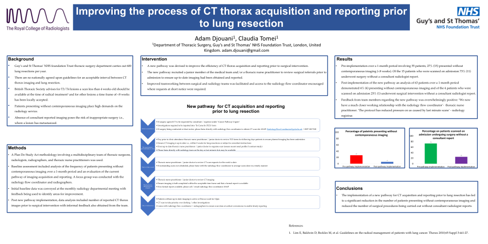
•A new pathway was devised to improve the efficiency of CT thorax acquisition and reporting prior to surgical intervention.
•The new pathway recruited a junior member of the medical team and/or a thoracic nurse practitioner to review surgical referrals prior to admission to ensure up-to-date imaging had been obtained and reported.
•Improved teamworking between surgical and radiology teams was facilitated and access to the radiology flow coordinator encouraged where requests at short notice were required.
Results
•Pre-implementation over a 1-month period involving 55 patients, 27% (15) presented without contemporaneous imaging (<8 weeks). Of the 15 patients who were scanned on admission 73% (11) underwent surgery without a consultant radiologist report.
•Post-implementation of the new pathway an analysis of 63 patients over a 1-month period demonstrated 6% (4) presenting without contemporaneous imaging and of the 4 patients who were scanned on admission 25% (1) underwent surgical intervention without a consultant radiologist report.
•Feedback from team members regarding the new pathway was overwhelmingly positive: ‘We now have a much closer working relationship with the radiology flow coordinator’- thoracic nurse practitioner. ‘The protocol has reduced pressure on us caused by last minute scans’ - radiology registrar.
Conclusions
•The implementation of a new pathway for CT acquisition and reporting prior to lung resection has led to a significant reduction in the number of patients presenting without contemporaneous imaging and reduced the number of surgical procedures being carried out without consultant radiologist reports.