16 posters, 11 topics, 56 authors, 21 institutions
ePostersLive by SciGen Technologies S.A. All rights reserved.
Clinical Radiology Audit ePoster Competition 2024
22 NOVEMBER, 2024 | LONDON, UK
88
Analysing Breast Density Reporting and Standardisation in Breast Mammogram Reports
Poster Presenter
Part of Topic
Breast
Introduction
Breast mammography aids in breast cancer detection as a 1st line investigation, with a primary benefit of reducing mortality due to its high accuracy [1]. However, dense breast tissue remains a big obstacle, increasing the risk of missed cancers and false reassurance due to hidden tumours [2]. Reporting breast density in mammography results provides patients and healthcare providers with insight into the likelihood of cancer detection based on
parenchymal patterns[3].
Aims
(i)reinforce the reporting of breast density in mammogram reports and the standardisation of breast density report using a common template.
(ii)ensure reporter includes documentation of previous mammograms availability at time of reporting.
(i)reinforce the reporting of breast density in mammogram reports and the standardisation of breast density report using a common template.
(ii)ensure reporter includes documentation of previous mammograms availability at time of reporting.
(iii)ensure at least 2-dimension measurement of lesion size is given.
(iv)ensure indication of mammogram was documented.
Standards
For breast density reporting, the American College of Radiology (ACR) Breast Imaging-Reporting and Data Systemic (BI-RADS) Breast Density Composition is used. Alphabetical A-D is used to described breast that are entirely fatty to entirely dense [4].
For standardisation of mammogram reports, both the Royal College of Radiologists (RCR) Breast Group breast imaging classification from 1-5 to describe normal to malignant findings [5] and ACR Breast Mammogram Reporting Guideline are used for mammogram report structure[4].
Report structure must include indication, reference to past mammogram, breast density, and clear description of important findings such as mass described in the report must be measured in 2 dimensions.
Methods
A retrospective analysis at Buckinghamshire Healthcare NHS Trust's Breast Imaging Department reviewed 790 mammogram reports from May to July 2023.
A re-audit was carried out from April to July 2024 involving 783 reports.
Inclusion criterion is mammograms from 2 week wait symptomatic clinics, excluding marker insertions and non-diagnostic mammograms.
Discussion
Breast Density
Report Structure
Breast Density
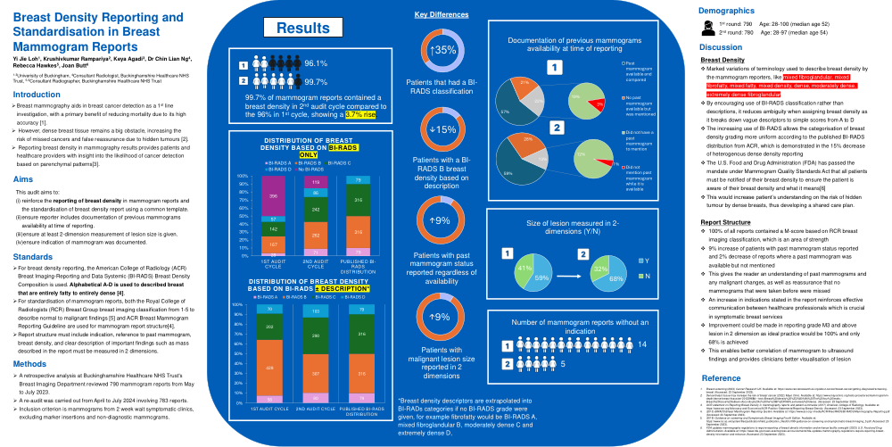
Marked variations of terminology used to describe breast density by the mammogram reporters, like mixed fibroglandular, mixed fibrofatty, mixed fatty, mixed density, dense, moderately dense, extremely dense fibroglandular
By encouraging use of BI-RADS classification rather than descriptions, it reduces ambiguity when assigning breast density as it breaks down vague descriptors to simple scores from A to D
The increasing use of BI-RADS allows the categorisation of breast density grading more uniform according to the published BI-RADS distribution from ACR, which is demonstrated in the 15% decrease of heterogenous dense density reporting
The U.S. Food and Drug Administration (FDA) has passed the mandate under Mammogram Quality Standards Act that all patients must be notified of their breast density to ensure the patient is aware of their breast density and what it means[6]
This would increase patient’s understanding on the risk of hidden tumour by dense breasts, thus developing a shared care plan.
Report Structure
100% of all reports contained a M-score based on RCR breast imaging classification, which is an area of strength
9% increase of patients with past mammogram status reported and 2% decrease of reports where a past mammogram was available but not mentioned
This gives the reader an understanding of past mammograms and any malignant changes, as well as reassurance that no mammograms that were taken before were missed
vAn increase in indications stated in the report reinforces effective communication between healthcare professionals which is crucial in symptomatic breast services
Improvement could be made in reporting grade M3 and above lesion in 2 dimension as ideal practice would be 100% and only 68% is achieved
This enables better correlation of mammogram to ultrasound findings and provides clinicians better visualisation of lesionv