16 posters, 11 topics, 56 authors, 21 institutions
ePostersLive by SciGen Technologies S.A. All rights reserved.
Clinical Radiology Audit ePoster Competition 2024
22 NOVEMBER, 2024 | LONDON, UK
82
From Protocol to Patient: Highlighting the Challenges of Translating CT Perfusion Standard Procedures into Effective Practice.
Poster Presenter
Authors
Affiliations
Part of Topic
Neuroradiology
From Protocol To Patient: Highlighting the Challenges of Translating CT Perfusion
Standard Procedures into Effective Practice
W. Southwell, V. Reay, F. Sheikh. University Hospital Southampton
|
INTRODUCTION Urgent CT perfusion is utilised to evaluate the perfusion of brain parenchyma in patients with acute stroke and reversible ischemia. In CT perfusion, the scan coverage is limited; therefore, the scan box must be positioned accurately. Inadequate positioning of the scan box has been iden0fied as a common issue in our Neuroradiology department. Consequently, an audit of CT Perfusion scan coverage was initiated in July 2022. |
|
STANDARD According to local guidelines, the scan should start at the floor of the pituitary fossa, unless clinical indications suggest otherwise. If there is any uncertainty, radiographers are advised to contact neuroradiology. |
|
METHOD Using a CRIS search, we identified all patients who underwent a CT Perfusion scan during two six-month periods (1st Audit: July to December 2022, 2nd Audit: August 2023 to January 2024). Upon retrospective review, the positioning of the inferior line of the scan box was categorised as follows: 'ACF floor/ sella', 'superior clivus', 'mid clivus', 'inferior clivus', and 'C1'. The patient's position on the table was also noted. Any documented deviations from the standard protocol were recorded. |
RESULTS
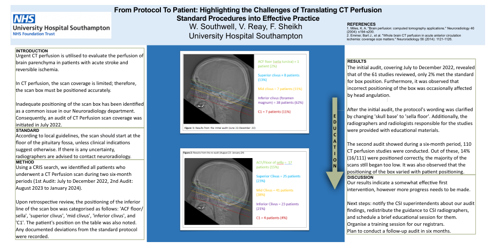
The initial audit, covering July to December 2022, revealed that of the 61 studies reviewed, only 2% met the standard for box position. Furthermore, it was observed that incorrect positioning of the box was occasionally affected by head angulation.
After the initial audit, the protocol's wording was clarified by changing 'skull base' to 'sella floor'. Additionally, the radiographers and radiologists responsible for the studies were provided with educational materials.
The second audit showed during a six-month period, 110 CT perfusion studies were conducted. Out of these, 14% (16/111) were positioned correctly, the majority of the scans still began too low. It was also observed that the positioning of the box varied with patient positioning.
DISCUSSION
Our results indicate a somewhat effective first intervention, however more progress needs to be made.
Next steps: notify the CSI superintendents about our audit findings, redistribute the guidance to CSI radiographers, and schedule a brief educational session for them. Organise a training session for our registrars.
Plan to conduct a follow-up audit in six months.
REFERENCES
1. Miles, K. A. "Brain perfusion: computed tomography applications." Neuroradiology 46 (2004): s194-s200.
2. Emmer, Bart J., et al. "Whole brain CT perfusion in acute anterior circulation ischemia: coverage size matters." Neuroradiology 56 (2014): 1121-1126.