16 posters, 11 topics, 56 authors, 21 institutions
ePostersLive by SciGen Technologies S.A. All rights reserved.
Clinical Radiology Audit ePoster Competition 2024
22 NOVEMBER, 2024 | LONDON, UK
77
<p>HOW GOOD ARE WE AT REPORTING VARICOCELES?:</p> <p>An Audit on Ultrasound evaluation and grading of varicoceles in compliance with European Society of Urogenital Radiology guidelines</p>
Poster Presenter
Authors
Affiliations
Part of Topic
Ultrasound
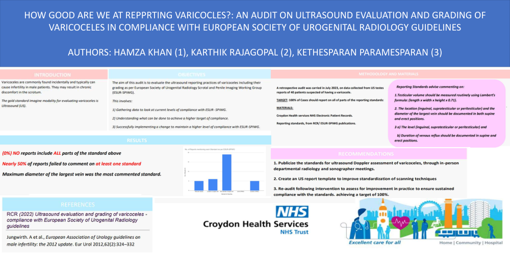
HOW GOOD ARE WE AT REPORTING VARICOCELES?:
An Audit on Ultrasound evaluation and grading of varicoceles in compliance with European Society of Urogenital Radiology guidelines
Authors: H, Khan (1), K. Rajagopal (2), K. Paramesparan (3).
Objectives:
The aim of this audit is to evaluate the ultrasound reporting practices of varicoceles including their grading as per European Society of Urogenital Radiology Scrotal and Penile Imaging Working Group (ESUR-SPIWG).
This involves:
1) Gathering data to look at current levels of compliance with ESUR- SPIWG.
2) Understanding what can be done to achieve a higher target of compliance.
3) Successfully implementing a change to maintain a higher level of compliance with ESUR-SPIWG.
Materials:
Croydon Health services NHS Electronic Patient Records.
Reporting standards, from RCR/ ESUR-SPIWG guidelines.
Methods:
A retrospective audit was carried on data collected from US reports of 40 patients in July 2023. Reporting standards advise including:
1. Testicular volume should be measured routinely using Lambert's formula: (length x width x height x 0.71).
2. The location (inguinal, supratesticular or peritesticular) and the diameter of the largest vein in both supine and erect positions.
3. The level (inguinal, supratesticular or peritesticular) and the duration of venous reflux in supine and erect positions.
Target: 100% of ultrasound reports should comment on all standards above.
Results: No reports commented on all standards above.
Conclusion:
Recommendations to improve compliance include:
1. Publicize the standards for ultrasound varicocele reporting, through in-person departmental radiology meetings and dissemination of written material to radiologists and sonographers.
2. Create a structured report template for use during electronic report transcription, in order to improve standardization of reporting items.
3. Re-audit 4-6 months after the intervention, to assess for improvement in practice. Continue the audit spiral, to ensure sustained compliance with the standards