29 posters, 13 topics, 90 authors, 29 institutions
ePostersLive by SciGen Technologies S.A. All rights reserved.
Clinical Radiology Audit and Quality Improvement ePoster Competition 2025
18 NOVEMBER, 2025 | LONDON, UK
91
Reducing the Orbital, Intracranial, and Overall Radiation Dose in Breast Cancer Patients Undergoing Staging CT
Poster Presenter
Authors
Affiliations
Part of Topic
Radiation dose/IR(ME)R
Reducing the Orbital, Intracranial, and Overall Radiation Dose in Breast Cancer Patients Undergoing Staging CT
P Nandra, N Motahariasl, W Teh, LNWUH NHS Trust
Introduction
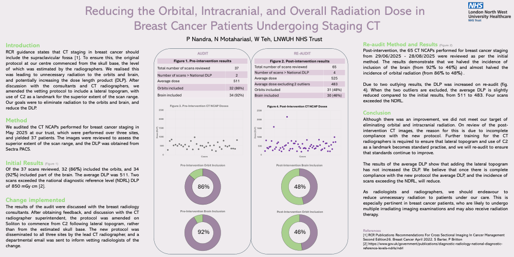
RCR guidance states that CT staging in breast cancer should include the supraclavicular fossa [1]. To ensure this, the original protocol at our centre commenced from the skull base, the level of which was estimated by the radiographers. We realised this was leading to unnecessary radiation to the orbits and brain, and potentially increasing the dose length product (DLP). After discussion with the consultants and CT radiographers, we amended the vetting protocol to include a lateral topogram, with use of C2 as a landmark for the superior extent of the scan range.
Our goals were to eliminate radiation to the orbits and brain, andreduce the DLP.
Method
We audited the CT NCAPs performed for breast cancer staging in May 2025 at our trust, which were performed over three sites, and yielded 37 patients. The images were reviewed to assess the superior extent of the scan range, and the DLP was obtained from Sectra PACS.
Initial Results(Figure 1)
Of the 37 scans reviewed, 32 (86%) included the orbits, and 34 (92%) included part of the brain. The average DLP was 511. Two scans exceeded the national diagnostic reference level (NDRL) DLP of 850 mGy cm [2].
Change implemented
The results of the audit were discussed with the breast radiology consultants. After obtaining feedback, and discussion with the CT radiographer superintendent, the protocol was amended on Soliton to commence from C2 following lateral topogram, rather than from the estimated skull base. The new protocol was disseminated to all three sites by the lead CT radiographer, and a departmental email was sent to inform vetting radiologists of the change.
Re-audit Method and Results (Figure 2)
Post-intervention, the 65 CT NCAPs performed for breast cancer staging from 29/06/2025 - 28/08/2025 were reviewed as per the initial method. The results demonstrate that we halved the incidence of inclusion of the brain (from 92% to 46%) and almost halved the incidence of orbital radiation (from 86% to 48%) .
Due to two outlying results, the DLP was increased on re-audit (fig. 4). When the two outliers are excluded, the average DLP is slightly reduced compared to the initial results, from 511 to 483. Four scans exceeded the NDRL.
Conclusion
Although there was an improvement, we did not meet our target of eliminating orbital and intracranial radiation. On review of the post-intervention CT images, the reason for this is due to incomplete compliance with the new protocol. Further training for the CT radiographers is required to ensure that lateral topogram and use of C2 as a landmark becomes standard practise, and we will re-audit to ensure that standards continue to improve.
The results of the average DLP show that adding the lateral topogramhas not increased the DLP. We believe that once there is complete compliance with the new protocol the average DLP, and the incidence of scans exceeding the NDRL, will reduce.
As radiologists and radiographers, we should endeavour to reduce unnecessary radiation to patients under our care. This is especially pertinent in breast cancer patients, who are likely to undergo multiple irradiating imaging examinations and may also receive radiation therapy.
References
[1] RCR Publications Recommendations For Cross Sectional Imaging In Cancer Management Second Edition26. Breast Cancer April 2022. S Barter, P Britton
[2] https://www.gov.uk/government/publications/diagnostic-radiology-national-diagnostic-reference-levels-ndrls/ndrl