29 posters, 13 topics, 90 authors, 29 institutions
ePostersLive by SciGen Technologies S.A. All rights reserved.
Clinical Radiology Audit and Quality Improvement ePoster Competition 2025
18 NOVEMBER, 2025 | LONDON, UK

90
Chest radiographs and ultrasound for the assessment of chest wall abnormalities in paediatrics.
Poster Presenter
Authors
Affiliations
Part of Topic
Paediatrics
Chest radiographs and ultrasound for the assessment of chest wall abnormalities in paediatrics
Dr Christopher Barrett (Junior Clinical Fellow), Ms Stephanie French (Specialist Paediatric Radiographer), Dr Mark Phillips (Consultant Paediatric Radiologist)
Leeds Teaching Hospitals NHS Trust
Background
GPs commonly refer children for chest X-rays (CXRs) for assessment of chest wall
abnormalities. Trust guidelines for Leeds Teaching Hospitals NHS Trust (LTHT)
advise:
• ‘Thoracic cage anomaly’ is a valid indication for paediatric CXR.
• Lateral views should be included.
• Ultrasounds (USS) should be considered to aid diagnosis.
This audit evaluated:
• Guideline adherence
• Whether imaging results were clinically actionable.
RCR Standards (2018) emphasise the importance of actionable imaging that
guides diagnosis and next steps in patient care. This audit
assessed appropriateness and impact of imaging in this context, using the
principle of actionable reporting as a benchmark.
Audit Standards
• Lateral views should be performed for CXRs investigating chest wall
abnormality.
• USS should be performed, especially where abnormalities are likely related to
costal cartilage concern.
• Imaging should provide clinically actionable information that influences
diagnosis or management
Methodology
A retrospective CRIS search identified all GP-requested CXRs for patients under
16 years across the calendar year of 2023, where the referral mentioned chest
wall abnormality (keywords : lump, deformity, asymmetry, prominence).
For each case, we recorded whether:
• CXR was normal or abnormal
• Abnormal findings were relevant to the clinical question or incidental
• Lateral view was performed
• USS was performed
• Imaging findings altered patient management (referral, treatment, follow-up)
Results
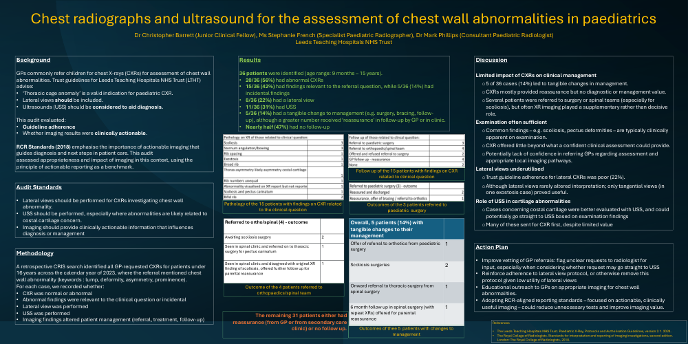
• 36 patients were identified (age range: 9 months – 15 years).
• 20/36 (56%) had abnormal CXRs
• 15/36 (42%) had findings relevant to the referral question, while 5/36 (14%) had
incidental findings
• 8/36 (22%) had a lateral view
• 11/36 (31%) had USS
• 5/36 (14%) had a tangible change to management (e.g. surgery, bracing, follow-
up), although a greater number received ‘reassurance’ in follow-up by GP or in clinic.
• Nearly half (47%) had no follow-up
The remaining 31 patients either had
reassurance (from GP or from secondary care
clinic) or no follow up.
Discussion
Limited impact of CXRs on clinical management
o 5 of 36 cases (14%) led to tangible changes in management.
o CXRs mostly provided reassurance but no diagnostic or management value.
o Several patients were referred to surgery or spinal teams (especially for
scoliosis), but often XR imaging played a supplementary rather than decisive
role.
Examination often sufficient
o Common findings – e.g. scoliosis, pectus deformities – are typically clinically
apparent on examination.
o CXR offered little beyond what a confident clinical assessment could provide.
o Potentially lack of confidence in referring GPs regarding assessment and
appropriate local imaging pathways.
Lateral views underutilised
o Trust guideline adherence for lateral CXRs was poor (22%).
o Although lateral views rarely altered interpretation; only tangential views (in
one exostosis case) proved useful.
Role of USS in cartilage abnormalities
o Cases concerning costal cartilage were better evaluated with USS, and could
potentially go straight to USS based on examination findings
o Many of these sent for CXR first, despite limited value
Action Plan
• Improve vetting of GP referrals: flag unclear requests to radiologist for
input, especially when considering whether request may go straight to USS
• Reinforce adherence to lateral view protocol, or otherwise remove this
protocol given low utility of lateral views
• Educational outreach to GPs on appropriate imaging for chest wall
abnormalities.
• Adopting RCR-aligned reporting standards – focused on actionable, clinically
useful imaging – could reduce unnecessary tests and improve imaging value.
References
• The Leeds Teaching Hospitals NHS Trust. Paediatric X-Ray, Protocols and Authorisation Guidelines, version 2.1. 2024.
• The Royal College of Radiologists. Standards for interpretation and reporting of imaging investigations, second edition.
London: The Royal College of Radiologists, 2018.