372 posters, 1 audios, 13 topics, 29 sessions, 1,035 authors, 449 institutions
ePostersLive by SciGen Technologies S.A. All rights reserved.
51st Annual Regional Anesthesiology and Acute Pain Medicine Meeting
April 16 - 18, 2026 | Phoenix, Arizona

2318754
Intrinsic angles and geometric modeling for thoracic epidurals: best approach analysis and prediction of LOR
Poster Presenter
Authors
Affiliations
Part of Topic
Scientific Abstracts > Regional Anesthesia
Intro:
•In addition to anatomical challenges encountered during placement, inadvertent advancement beyond the intended interspace may occur during epidural needle insertion.
•Traditional thoracic epidural techniques often ignore 3D constraints.
•We propose that each thoracic level possesses intrinsic angles of needle entry, and that aligning the needle trajectory with this geometry may improve overall efficiency, accuracy, and concordant analgesic coverage.
Methods:
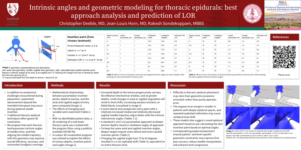
•Mathematical relationships between paramedian insertion points, depth to lamina, and the axial and sagittal angles of entry were computed (Image 1).
•The effects of changing each variable were examined (Tables 1-3).
•With no identifiable patient data, a 3D rendering of a mid-lower thoracic spine was created with the program Slicer using a publicly available DICOM file.
•A custom 3D visualization program was utilized to explore the effects of various depths, insertion points and angles (Image 2).
Results:
•Increased depth to the lamina progressively narrows the effective interlaminar window, and at greater depths, small changes in axial or sagittal angulation will result in level shifts, increasing osseous contacts, or failed blocks (visualized in image 2)
•A more lateral and caudad skin entry point with a relatively increased medial and relatively decreased sagittal needle trajectory aligns better with the intrinsic interlaminar angles (Tables 1-2).
•A standard 1 cm/1 cm paramedian approach at deeper laminar depths results in shallower angles of approach
•To keep the same axial and sagittal insertion angles, deeper targets require more lateral and more caudad insertion points (Table 2).
•Changing the sagittal angle from 75 to 55 degrees resulted in a 2 cm cephalad shift (Table 3), equivalent to an entire thoracic level.
Discussion:
•Difficulty in thoracic epidural placement may stem from geometric/anatomic mismatch rather than purely operator error.
•The angular error margin is smaller in patients with deeper epidural spaces, and repeated cephalad redirection may cause vertebral level shift.
•These models also suggest a novel epidural approach based on pre-calculating the skin insertion point based on optimal angles.
•Conceptualizing epidural placement around patient- and level-specific geometric constraints may improve first-pass success, reduce needle manipulation, and enhance level congruence.