372 posters, 1 audios, 13 topics, 29 sessions, 1,035 authors, 449 institutions
ePostersLive by SciGen Technologies S.A. All rights reserved.
51st Annual Regional Anesthesiology and Acute Pain Medicine Meeting
April 16 - 18, 2026 | Phoenix, Arizona

2316185
“Where Did It Go?” to “Now I See It”: Contrast Imaging Improves Resident Mental Models of Local Anesthetic Spread
Poster Presenter
Authors
Affiliations
Part of Topic
Scientific Abstracts > Education
Contrast-Confirmed Injectate Spread as a Teaching Tool for Regional Anesthesia Residents: A Pediatric Oncology Experience
Introduction
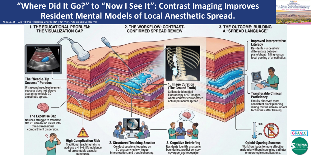
A major challenge in regional anesthesia training is translating ultrasound-guided needle placement into a reliable understanding of injectate spread, particularly in fascial plane blocks, where sonographic appearance may not reflect true clinical coverage. Contrast-assisted fluoroscopy or CT may provide imaging “ground truth,” helping trainees connect sonoanatomy with three-dimensional dispersion. We describe a resident-focused teaching workflow based on contrast-confirmed images from two pediatric oncology pain cases.
Materials and Methods
At a pediatric oncology referral hospital in Brazil, de-identified contrast images from clinically indicated radiologic studies were curated for a structured teaching session including 3D anatomy review, image interpretation, and troubleshooting. Twenty anesthesiology residents (16 PGY-2, 4 PGY-3) participated. Written guardian consent for educational use of de-identified images was obtained, and all protected health information was removed. Per institutional policy, the project was IRB-exempt.
Two cases were used:
Case 1: continuous costoclavicular brachial plexus catheter for upper-limb disarticulation; fluoroscopy with iodinated contrast confirmed perineural spread.
Case 2: a 17-year-old undergoing liver tumor ablation received QL type 3 and lumbar ESP blocks; CT with contrast demonstrated injectate dispersion. Off-label perineural use of iodinated contrast was documented.
Results
Contrast imaging helped residents distinguish expected from inadequate spread patterns, including sheath/plane filling versus focal pooling. During structured debriefing, trainees identified relevant anatomic boundaries, predicted likely sensory coverage, and discussed troubleshooting strategies such as malposition and incomplete plane opening. Residents consistently reported improved understanding of injectate dispersion after reviewing fluoroscopy and CT images. Faculty also observed more consistent spread interpretation and block planning in subsequent routine ultrasound-guided practice. In both index cases, analgesia was effective and opioid-sparing, with no contrast-related adverse events, neurologic deficits, or catheter complications.
Discussion
Contrast-confirmed imaging may be a high-yield educational adjunct for teaching injectate spread in continuous plexus catheters and fascial plane blocks. Because contrast and radiation add cost and risk, this strategy should remain limited to carefully selected cases in which imaging is clinically justified and de-identified images can be reused for broader teaching. Future work should quantify learning gains and correlate them with procedural performance and patient-centered outcomes.
References
-
Diwan S, Sethi D, Sancheti P, Nair A. Braz J Anesthesiol. 2023;73(3):347-350.
-
Adhikary SD, El-Boghdadly K, Nasralah Z, et al. Anaesthesia. 2017;72(1):73-79.