343 posters, 5 videos, 2 audios, 18 topics, 33 sessions, 263 authors, 202 institutions
ePostersLive by SciGen Technologies S.A. All rights reserved.
24th Annual Pain Medicine Meeting 2025
November 13-15, 2025| JW Marriott Austin, Austin, Texas

2197911
Beyond Radiculopathy: A Missed Sciatic Nerve Sheath Tumor Mimicking Radicular Pain
Poster Presenter
Authors
Affiliations
Part of Topic
Medically Challenging Cases
Beyond Radiculopathy: A Missed Sciatic Nerve Sheath Tumor Mimicking Radicular Pain
Antoine Sioufi, MD,Ramsis Ghaly, MD, Taruna Penmetcha, MD
John Stroger Hospital of Cook County, Chicago, IL
Introduction: Lumbar radiculopathy commonly causing lower extremity painis often attributed to degenerative spinal pathology [2]. However, when patientsfail to respond to conventional treatments, it is imperative to reassess the diagnosis and considerless commonetiologies. This case highlights a patient initially diagnosed and treated for lumbar radiculopathy who continued to experience progressive symptoms despite multiple interventional and conservative therapies. Further investigationultimately revealed a sciatic nerve tumor as the underlying cause of the persistent pain.
Materials and methods: As the case report is devoid of patient identifiable information, it is exempt from IRB review requirements as per Cook County Health policy. Patient informed consent was obtained for submission of the case report.
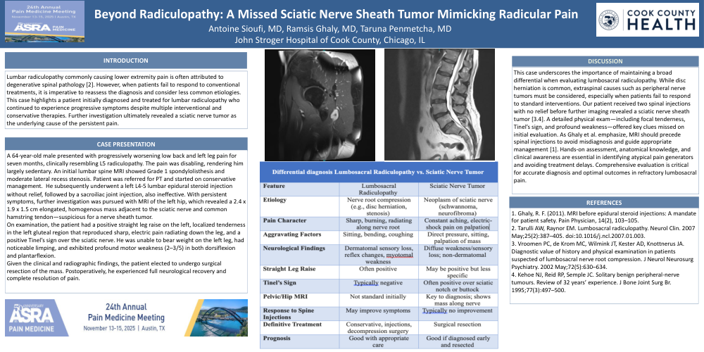
Case report:A 64-year-old male presented with progressively worsening low back and left leg pain for seven months, clinically resembling L5 radiculopathy. The pain was disabling, rendering him largely sedentary. An initial lumbar spine MRI showed Grade 1 spondylolisthesis and moderate lateral recess stenosis. Patient was referred for PT and started on conservative management. He subsequentlyunderwent a left L4-5 lumbar epidural steroid injection without relief, followed by a sacroiliac joint injection, also ineffective. With persistent symptoms, further investigation was pursued with MRI of the left hip, which revealed a 2.4 x 1.9 x 1.5 cm elongated, homogenous mass adjacent to the sciatic nerve and common hamstring tendon—suspicious for a nerve sheath tumor.
On examination, the patient had a positive straight leg raise on the left, localized tenderness in the left gluteal region that reproduced sharp, electric pain radiating down the leg, and a positive Tinel’s sign over the sciatic nerve. He was unable to bear weight on the left leg, had noticeable limping, and exhibited profound motor weakness (2–3/5) in both dorsiflexion and plantarflexion.
Given the clinical and radiographic findings, the patient elected to undergo surgical resection of the mass. Postoperatively, he experienced full neurological recovery and complete resolution of pain.
Discussion:This case underscores the importance of maintaining a broad differential when evaluating lumbosacral radiculopathy. While disc herniation is common, extraspinal causes such as peripheral nerve tumors must be considered, especially when patients fail to respond to standard interventions. Our patient received two spinal injections with no relief before further imaging revealed a sciatic nerve sheath tumor [3.4]. A detailed physical exam—including focal tenderness, Tinel’s sign, and profound weakness—offered key clues missed on initial evaluation. As Ghaly et al. emphasize, MRI should precede spinal injections to avoid misdiagnosis and guide appropriate management [1]. Hands-on assessment, anatomical knowledge, and clinical awareness are essential in identifying atypical pain generators and avoiding treatment delays. Comprehensive evaluation is critical for accurate diagnosis and optimal outcomes in refractory lumbosacral pain.
Reduced to this due to 75 word count
This case highlights the need for a broad differential when evaluating lumbosacral radiculopathy. Though disc herniation is common, extraspinal causes like nerve sheath tumors must be considered when standard treatments fail. Our patient had no relief from two spinal injections before MRI revealed a sciatic nerve tumor [3,4]. As Ghaly et al. emphasize, MRI should precede injections [1]. Detailed exams, including Tinel’s sign and weakness, are essential for identifying atypical pain sources and preventing delays.
References:
1. Ghaly, R. F. (2011). MRI before epidural steroid injections: A mandate for patient safety. Pain Physician, 14(2), 103–105.
2. Tarulli AW, Raynor EM. Lumbosacral radiculopathy. Neurol Clin. 2007 May;25(2):387–405. doi:10.1016/j.ncl.2007.01.003.
3. Vroomen PC, de Krom MC, Wilmink JT, Kester AD, Knottnerus JA. Diagnostic value of history and physical examination in patients suspected of lumbosacral nerve root compression. J Neurol Neurosurg Psychiatry. 2002 May;72(5):630–634.
4. Kehoe NJ, Reid RP, Semple JC. Solitary benign peripheral-nerve tumours. Review of 32 years’ experience. J Bone Joint Surg Br. 1995;77(3):497–500.