343 posters, 5 videos, 2 audios, 18 topics, 33 sessions, 263 authors, 202 institutions
ePostersLive by SciGen Technologies S.A. All rights reserved.
24th Annual Pain Medicine Meeting 2025
November 13-15, 2025| JW Marriott Austin, Austin, Texas

2183944
Ultrasound-Guided PRP for Interspinous Ligament Defect and Bursitis After Clay-Shoveler’s Fracture in Hypermobile Teen
Poster Presenter
Authors
Affiliations
Part of Topic
Medically Challenging Cases
Ultrasound-Guided PRP for Interspinous Ligament Defect and Bursitis After Clay-Shoveler’s Fracture in Hypermobile Teen
Alan Shain, MD,1 Jeremy R. Benhamroun-Zbili, DO,2
1 Long Island Jewish Forest Hills Hospital, Queens, New York, USA.
2 Northwell Greenwich Village Hospital, New York, New York, USA.
BACKGROUND AND PURPOSE: Clay-shoveler’s fractures and interspinous ligament injuries in pediatric patients are rarely atraumatic even in the higher-risk connective tissue disorder population.1,2 Ultrasound-guided Platelet-Rich Plasma (PRP) has limited but emerging evidence as a targeted treatment for refractory interspinous ligament injuries.3,4 We present a 16-year-old male with suspected Ehlers-Danlos Syndrome (EDS) who developed atraumatic T2–T4 cervicothoracic instability refractory to C7 Clay-shoveler’s fracture repair, improving with PRP prior to an eventual Amplified Musculoskeletal Pain Syndrome (AMPS) diagnosis.
SETTING: Outpatient Physical Medicine and Rehabilitation Clinic
MATERIALS & METHODS: This is a retrospective case report of a 16-year-old male with benign hypermobility syndrome, family history of Ehlers-Danlos syndrome, and prior surgically-treated C7 spinous process avulsion. A post-operative MRI following C7 spinous process resection showed mild C7–T2 degenerative changes and early interspinous bursitis. Nine months post-resection, another MRI revealed a T3 supraspinous ligament avulsion and T3–T4 interspinous sprains. The patient underwent anatomically-guided T1–T2 trigger point injections, followed by a series of ultrasound-guided in-plane injections: a T3 adventitious bursa steroid injection, a T3–T5 interspinous ligaments and bursae prolotherapy/steroid injection, and then a T2–T4 PRP (5 mL PRP + 15 mL PPP) injection. He received a thoracolumbosacral orthosis (TLSO) and participated in an eight-week interdisciplinary rehabilitation program. PRP was used off-label per IRB policy and standard practice. It is considered experimental by the FDA. Patient informed consent was obtained. This de-identified case report is exempt from IRB-review per Northwell Health policy.
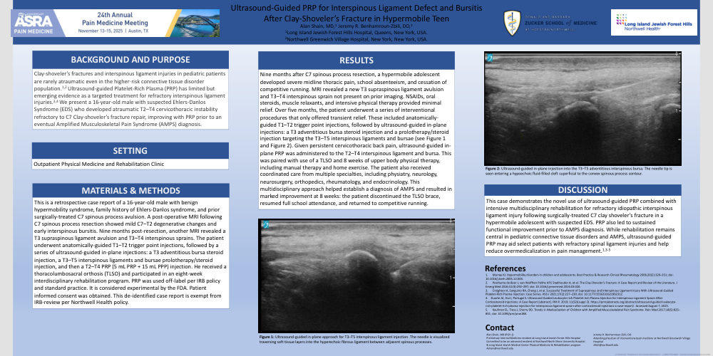
RESULTS: Nine months after C7 spinous process resection, a hypermobile adolescent developed severe midline thoracic pain, school absenteeism, and cessation of competitive running. MRI revealed a new T3 supraspinous ligament avulsion and T3–T4 interspinous sprain not present on prior imaging. NSAIDs, oral steroids, muscle relaxants, and intensive physical therapy provided minimal relief. Over five months, the patient underwent a series of interventional procedures that only offered transient relief. These included anatomically-guided T1–T2 trigger point injections, followed by ultrasound-guided in-plane injections: a T3 adventitious bursa steroid injection and a prolotherapy/steroid injection targeting the T3–T5 interspinous ligaments and bursae (see Figure 1 and Figure 2). Given persistent cervicothoracic back pain, ultrasound-guided in-plane PRP was administered to the T2–T4 interspinous ligament and bursa. This was paired with use of a TSLO and 8 weeks of upper body physical therapy, including manual therapy and home exercise. The patient also received coordinated care from multiple specialties, including physiatry, neurology, neurosurgery, orthopedics, rheumatology, and endocrinology. This multidisciplinary approach helped establish a diagnosis of AMPS and resulted in marked improvement at 8 weeks: the patient discontinued the TLSO brace, resumed full school attendance, and returned to competitive running.
DISCUSSION: This case demonstrates the novel use of ultrasound-guided PRP combined with intensive multidisciplinary rehabilitation for refractory idiopathic interspinous ligament injury following surgically-treated C7 clay shoveler’s fracture in a hypermobile adolescent with suspected EDS. PRP also led to sustained functional improvement prior to AMPS diagnosis. While rehabilitation remains central in pediatric connective tissue disorders and AMPS, ultrasound-guided PRP may aid select patients with refractory spinal ligament injuries and help reduce overmedicalization in pain management.1,3-5
References
1. Murray KJ. Hypermobility disorders in children and adolescents. Best Practice & Research Clinical Rheumatology 2006;20(2):329–351; doi: 10.1016/j.berh.2005.12.003.
2. Posthuma de Boer J, van Wulfften Palthe AFY, Stadhouder A, et al. The Clay Shoveler’s Fracture: A Case Report and Review of the Literature. J EmergMed 2016;51(3):292–297; doi: 10.1016/j.jemermed.2016.03.020.
3. Creighton A, Sanguino RA, Cheng J, et al. Successful Treatment of Supraspinous and Interspinous Ligament Injury With Ultrasound-Guided Platelet-Rich Plasma Injection: Case Series. HSS J 2021;17(2):227–230; doi: 10.1177/1556331621992312.
4. Duarte AJ, Hui J, Portugal S. Ultrasound Guided Leukocyte-rich Platelet-rich Plasma Injection for Interspinous Ligament Sprain After Corticosteroid Injections: A Case Report [abstract]. PM R. 2019; 11(S2)(suppl 2). https://pmrjabstracts.org/abstract/ultrasound-guided-leukocyte-rich-platelet-rich-plasma-injection-for-interspinous-ligament-sprain-after-corticosteroid-injections-a-case-report/. Accessed August 7, 2025.
5. Kaufman EL, Tress J, Sherry DD. Trends in Medicalization of Children with Amplified Musculoskeletal Pain Syndrome. Pain Med 2017;18(5):825–831; doi: 10.1093/pm/pnw188.