126 posters, 2 topics, 27 sessions, 324 authors, 82 institutions
ePostersLive by SciGen Technologies S.A. All rights reserved.
2025 ACP Annual Session
October 22 - 25, 2025 | New Orleans, US

30
Novel 3D-Printed Palatal Expansion Device Optimizing Full-Mouth Restorative Protocols
Poster Presenter
Authors
Affiliations
Part of Topic
Case Presentation
A Novel 3D Printed Fully-guided Palatal Expansion Device in Support of Full Mouth Rehabilitation
Matthew J. Turner DDS, LCDR/ DC/ USN; Barry E. Peterson, DDS, MS, CAPT/DC/USN; Timothy P. Daudelin, DDS, MS, FACP, LCDR/DC/USN; Stacy L. Yu, DDS, FACP, CDR/DC/USN
Naval Postgraduate Dental School, Uniformed Services University of the Health Sciences, Walter Reed National Military Medical Center, Bethesda, MD
Correction of maxillary transverse deficiency in adults traditionally involves invasive surgical procedures such as surgically assisted rapid palatal expansion (SARPE) or segmental Le Fort I osteotomy.1 However, the integration of computer-aided design and manufacturing (CAD/CAM) technologies has enabled the use of targeted mini-implant-assisted rapid palatal expansion (MARPE) appliances as a less invasive alternative with predictable outcomes.2 Minimizing surgical complications and reducing post-operative healing time are critical considerations during treatment planning.
Figure 1: Patient presentation with maxillary transverse deficiency, forward functional shift, and collapsed occlusal vertical dimension (OVD) in need of a fixed full mouth rehabilitation.
To outline the planning, design, and implementation of a novel palatal expansion device aimed at correcting a bilateral reverse articulation with a forward functional shift and generalized tooth structure loss in a 44-year-old Turner Class I adult male as part of preparatory treatment prior to restorative rehabilitation with fixed restorations.
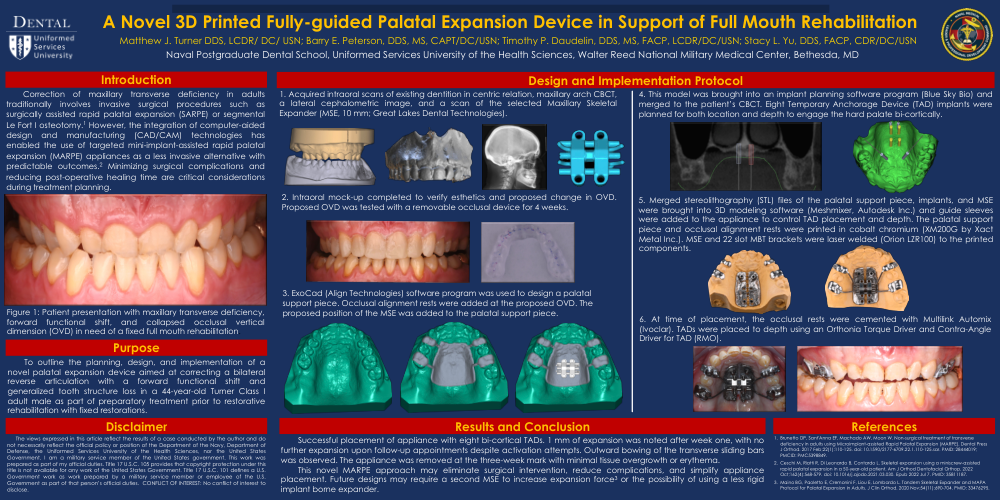
1. Acquired intraoral scans of existing dentition in centric relation, maxillary arch CBCT, a lateral cephalometric image, and a scan of the selected Maxillary Skeletal Expander (MSE, 10 mm; Great Lakes Dental Technologies).
2. Intraoral mock-up completed to verify esthetics and proposed change in OVD. Proposed OVD was tested with a removable occlusal device for 4 weeks.
3. ExoCad (Align Technologies) software program was used to design a palatal support piece. Occlusal alignment rests were added at the proposed OVD. The proposed position of the MSE was added to the palatal support piece.
4. This model was brought into an implant planning software program (Blue Sky Bio) and merged to the patient’s CBCT. Eight Temporary Anchorage Device (TAD) implants were planned for both location and depth to engage the hard palate bi-cortically.
5. Merged stereolithography (STL) files of the palatal support piece, implants, and MSE were brought into 3D modeling software (Meshmixer, Autodesk Inc.) and guide sleeves were added to the appliance to control TAD placement and depth. The palatal support piece and occlusal alignment rests were printed in cobalt chromium (XM200G by Xact Metal Inc.). MSE and 22 slot MBT brackets were laser welded (Orion LZR100) to the printed components.
6. At time of placement, the occlusal rests were cemented with Multilink Automix (Ivoclar). TADs were placed to depth using an Orthonia Torque Driver and Contra-Angle Driver for TAD (RMO).
Successful placement of appliance with eight bi-cortical TADs. 1 mm of expansion was noted after week one, with no further expansion upon follow-up appointments despite activation attempts. Outward bowing of the transverse sliding bars was observed. The appliance was removed at the three-week mark with minimal tissue overgrowth or erythema. This novel MARPE approach may eliminate surgical intervention, reduce complications, and simplify appliance placement. Future designs may require a second MSE to increase expansion force3 or the possibility of using a less rigid implant borne expander.