126 posters, 2 topics, 27 sessions, 324 authors, 82 institutions
ePostersLive by SciGen Technologies S.A. All rights reserved.
2025 ACP Annual Session
October 22 - 25, 2025 | New Orleans, US

123
Prosthodontic Rehabilitation of an 8-Year-Old Male with Ectodermal Dysplasia Using Flexible Partial Dentures: A Case Report
Poster Presenter
Authors
Affiliations
Part of Topic
Case Presentation
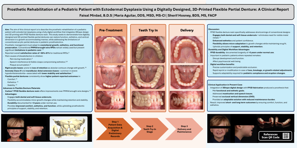
Prosthetic Rehabilitation of a Pediatric Patient with Ectodermal Dysplasia Using a Digitally Designed, 3D-Printed Flexible Partial Denture: A Clinical Report
Keywords: ectodermal dysplasia, pediatric prosthodontics, hypodontia/anodontia, digital dentures, 3Shape design, FP3D material, flexible partial denture, growth modification
Abstract
This clinical report details the prosthetic rehabilitation of an 8-year-old boy with ectodermal dysplasia, presenting with retained primary anterior teeth, erupting permanent central incisors, unerupted permanent canines, and absence of all other teeth. The patient exhibited a skeletal Class III relationship with a severe anterior crossbite. Treatment aimed to restore masticatory function and speech, preserve vertical dimension and arch integrity, and improve esthetics in a growth-accommodating manner. A fully digital workflow was implemented using 3Shape for design, followed by a physical teeth try-in to verify occlusion, esthetics, and comfort. The final appliance was fabricated using FP3D, an FDA-approved Class II flexible partial denture resin, noted for its high precision, excellent tooth retention, and durability beyond three years. This hybrid approach—digital design, printed try-in, and advanced polymer fabrication—aligns with current recommendations for early, minimally invasive prosthetic management in pediatric ED patients while enhancing efficiency, accuracy, and patient acceptance.
Introduction
Ectodermal dysplasias (ED) are a genetically diverse group of disorders first categorized by Freire-Maia in 19711 and reclassified in 2014 into at least 186 distinct syndromes.2 All ED conditions involve abnormal development of at least two ectoderm-derived structures, including hair, teeth, nails, and sweat glands, with an estimated global incidence of ~7/10,000 births. 3 The overall period prevalence for hypohidrotic ectodermal dysplasia (HED) in a large U.S. multicenter database was estimated at 2.99 per 100,000 individuals.4 The most common inheritance pattern is X-linked recessive, with expression more frequent in males.3
Oral manifestations are significant and include hypodontia or anodontia in both primary and permanent dentitions, abnormal eruption timing, reduced tooth size, conical or fused teeth, taurodontism, malposition, and abnormal spacing.5 These can impede jaw development and alveolar ridge formation. Genetic testing and counseling are encouraged to facilitate care planning and access to support resources.
Dental rehabilitation in ED requires a multidisciplinary approach (pediatric dentistry, orthodontics, prosthodontics, oral and maxillofacial surgery) to provide an age-appropriate dentition that supports mastication, facial development, speech, esthetics, and psychosocial well-being.6–8 Planning must consider age, psychosocial environment, residual dentition, oral hygiene, occlusal vertical dimension (OVD), bone availability, jaw growth, implant timing, maintenance needs, and cost.9,10 Preservation of existing dentition, especially primary teeth lacking successors, is critical to maintain alveolar bone, assist in prosthesis retention, and serve as orthodontic anchorage when appropriate.11
Early intervention typically involves composite restoration of malformed anterior teeth and interim removable prostheses to restore function and appearance. While effective, conventional acrylic partials require excellent home care and have well-documented limitations. A recent systematic review reported satisfaction rates with conventional RPDs ranging from ~50% to 81%, with common complaints including pain during mastication, compromised esthetics from visible clasps, speech interference, plaque retention, and poor adaptation to growth-related changes.12 Rigid acrylic bases are particularly prone to loss of retention as ridge contours evolve, and designs involving extensive tooth loss, Kennedy Class IV configurations, or mandibular distal extension bases often yield lower satisfaction. Metal clasp–retained designs, while mechanically retentive, may cause soft tissue irritation and detract from esthetics. Flexible materials, attachment-retained designs, and implant-assisted RPDs consistently score higher in comfort, esthetics, and stability.
The advent of Carbon® FP3D flexible denture resin enables a departure from the initial PMMA/wrought wire design concept, which relied on clasp engagement of dental undercuts. Instead, the denture base can now engage both dental and soft tissue undercuts, leveraging the material’s flexibility to accommodate minor growth-related changes while maintaining retention and stability. With documented durability exceeding 3 years under normal use, the advantages of a new design provides a snug, adaptive fit that addresses multiple shortcomings of traditional interim dentures—offering improved comfort, esthetics, and function while upholding core removable prosthodontic principles of support, stability, and retention.
Clinical Report
Patient and Diagnosis
An 8-year-old boy with hypohidrotic ED presented with difficulty chewing, speech concerns, and esthetic dissatisfaction. Clinical and radiographic evaluation revealed:
- Retained primary anterior teeth (#C, #H, #M, #N, #P, #Q, #R)
- Erupting permanent maxillary central incisors (#8, #9)
- Unerupted maxillary permanent canines (#6, #11)
- Complete absence of all other primary and permanent teeth
- Skeletal Class III relationship with severe anterior crossbite
- Narrow arches and reduced posterior alveolar height
Treatment objectives included restoring posterior support, maintaining OVD, improving esthetics, and delivering a prosthesis compatible with craniofacial growth.
Step 1 — Patient Radiographic Data Acquisition & Digital Preliminary Records
Radiographic examination, intraoral and extraoral photos were taken. Preliminary impressions obtained via intraoral scanning using a 3Shape Trios system to maximize comfort and avoid bulky trays. Digital files allowed precise visualization of arch form, occlusion, and space distribution, and served as the foundation for virtual tooth setup.
Step 2 — Physical Try-In Stage
A 3D-printed try-in of the proposed tooth arrangement was fabricated and delivered to verify occlusion, esthetics, speech, and patient comfort. This stage allowed approval from both clinician and guardian prior to definitive processing.
Step 3 — Final Fabrication Using FP3D Material
The final prosthesis was manufactured using Carbon® FP3D flexible denture resin. Advantages included:
- Durability exceeding 3 years under normal use
- High translucency for natural tissue blending
- Excellent mechanical retention of denture teeth
- Superior polishability compared to thermoplastics
- Low water sorption (1.82 µg/mm³) and solubility (0.01 µg/mm³)
- ISO 10993 biocompatibility compliance
Step 4 — Delivery and Maintenance
The FP3D flexible partial denture was inserted, ensuring even occlusion and smooth tissue adaptation. Written and verbal instructions were given regarding hygiene and insertion/removal techniques. Follow-ups were scheduled every 3 months to monitor growth and adjust retention as needed.
Discussion
Prosthetic management of pediatric ED patients presents unique challenges due to the need for ongoing adaptation to craniofacial growth, maintenance of esthetics, and preservation of oral function. Conventional PMMA/wrought wire RPDs, while widely used, have well-documented limitations in this context. The recent systematic review by Awawdeh et al. reported that although overall satisfaction rates for traditional RPDs ranged from ~50% to 81%, dissatisfaction often stemmed from factors directly relevant to pediatric care—pain during mastication, visible clasping compromising esthetics, interference with speech, and plaque accumulation. Rigid acrylic bases are particularly susceptible to retention loss as alveolar contours change, which is an unavoidable process during a child’s growth phase. Additionally, designs involving Kennedy Class IV or mandibular distal extension bases, both common in severe hypodontia/anodontia cases, were associated with lower stability and satisfaction.
These limitations underscore the need for alternative materials and retention strategies that can maintain function and comfort while accommodating anatomical changes. Flexible partial dentures have consistently shown higher patient-reported outcomes in comfort, esthetics, and stability compared to metal or rigid acrylic designs. The FP3D flexible denture resin used in this case offers several advantages that directly address the shortcomings highlighted in the literature. Its ability to engage both dental and soft tissue undercuts eliminates the need for visible metal clasps, enhancing esthetics and patient confidence. The material’s flexibility allows for micro-adaptation to growth-related changes, maintaining a snug, comfortable fit without compromising core prosthodontic principles of support, stability, and retention.
From a durability perspective, FP3D has documented longevity exceeding 3 years under normal use—an important consideration in pediatric cases where repeated remakes can be disruptive to development, function, and psychosocial well-being. The fully digital workflow also provides additional benefits: precise customization, predictable occlusal schemes, and the ability to rapidly reprint or modify the prosthesis in the event of loss or damage. This is particularly valuable in pediatric patients, where growth, eruption patterns, and compliance can necessitate timely prosthetic adjustments.
In the present case, the integration of 3Shape digital design with FP3D material fabrication allowed for a prosthesis that not only met functional and esthetic objectives but also aligned with the long-term needs of a growing child with ED. The resulting appliance effectively addressed the patient’s masticatory and speech concerns, preserved vertical dimension, and offered an adaptable solution with reduced maintenance burden—key factors in improving both short- and long-term treatment outcomes.
This case supports the incorporation of digitally designed, flexible-material prostheses such as FP3D into pediatric ED protocols, offering an evidence-based pathway to improve comfort, function, and esthetics while reducing the frequency of intervention during growth.
Table 1. Comparison of Literature-Identified Limitations of Conventional Interim RPDs and Advantages of FP3D Flexible Digital Designs
|
Feature |
Conventional Interim RPDs |
FP3D Flexible Digital Design |
|
Patient Satisfaction |
Satisfaction rates ~50–81%; lower in Kennedy Class IV, distal extension bases, or extensive tooth loss |
High comfort and esthetics; no visible clasps; adaptable to various edentulous configurations |
|
Retention & Stability |
Retention loss over time due to growth-related ridge changes; rigid base limits adaptation |
Engages both dental and soft tissue undercuts; flexibility maintains fit during minor anatomical changes |
|
Esthetics |
Visible metal clasps compromise appearance; lower esthetic ratings in anterior tooth loss cases |
No visible metal; high translucency blends with gingival tissues |
|
Comfort |
Rigid acrylic bases can cause soft tissue irritation and bulkiness |
Lightweight, flexible base reduces irritation and enhances comfort |
|
Speech |
Potential interference, particularly with rigid PMMA bases |
Close adaptation to palatal/lingual contours supports clear speech |
|
Hygiene |
Plaque retention around clasps; cleaning challenges |
Smooth, highly polishable surface facilitates cleaning; clasp-free design minimizes plaque traps |
|
Durability |
Susceptible to fracture and clasp distortion |
Documented durability exceeding 3 years under normal use; maintains mechanical retention of denture teeth |
|
Maintenance |
Multiple relines or adjustments often required during growth; analog workflow limits reproducibility |
Digital design allows rapid reprint or modification without full remake |
|
Adaptability to Growth |
Poor—requires frequent intervention to accommodate changing jaw morphology |
Flexibility accommodates minor growth changes while maintaining retention and stability |
Caption: Comparative summary of key limitations of conventional interim removable partial dentures (RPDs) as reported in a systematic review by Awawdeh et al. and the clinical advantages of FP3D flexible digital designs in pediatric prosthetic rehabilitation.
References
1. Freire-Maia N. Ectodermal Dysplasias. Hum Hered. 1971;21(4):309-312. doi:10.1159/000152419
2. Pagnan NAB, Visinoni ÁF. Update on ectodermal dysplasias clinical classification. Am J Med Genet A. 2014;164(10):2415-2423. doi:10.1002/ajmg.a.36616
3. Majmundar VD, Baxi K. Ectodermal Dysplasia. StatPearls. July 24, 2023. Accessed August 11, 2025. https://www.ncbi.nlm.nih.gov/books/NBK563130/
4. Butcher C, Abbott BM, Grange D, et al. Prevalence rates for ectodermal dysplasia syndromes. Am J Med Genet A. 2024;194(12):e63832. doi:10.1002/ajmg.a.63832
5. Míšová E, Kratochvilova L, Dostalova T, et al. Ectodermal dysplasia: important role of complex dental care in its interdisciplinary management. Eur J Paediatr Dent. 2022;23(2):140-146. doi:10.23804/ejpd.2022.23.02.12
6. Ioannidou-Marathiotou I, Kotsiomiti E, Gioka C. The Contribution of Orthodontics to the Prosthodontic Treatment of Ectodermal Dysplasia. J Am Dent Assoc. 2010;141(11):1340-1345. doi:10.14219/jada.archive.2010.0078
7. Yenisey M, Guler A, Unal U. Orthodontic and prosthodontic treatment of ectodermal dysplasia — a case report. Br Dent J. 2004;196(11):677-679. doi:10.1038/sj.bdj.4811344
8. Lypka M, Yarmand D, Burstein J, Tso V, Yamashita DD. Dental Implant Reconstruction in a Patient With Ectodermal Dysplasia Using Multiple Bone Grafting Techniques. J Oral Maxillofac Surg. 2008;66(6):1241-1244. doi:10.1016/j.joms.2008.01.004
9. Bergendal B, Bjerklin K, Bergendal T, Koch G. Dental Implant Therapy for a Child with X-linked Hypohidrotic Ectodermal Dysplasia— Three Decades of Managed Care. Int J Prosthodont. 2015;28(4):348-356. doi:10.11607/ijp.4242
10. Vergo TJ. Prosthodontics for pediatric patients with congenital/developmental orofacial anomalies: A long-term follow-up. J Prosthet Dent. 2001;86(4):342-347. doi:10.1067/mpr.2001.118877
11. Levander E, Malmgren O, Stenback K. Apical root resorption during orthodontic treatment of patients with multiple aplasia: a study of maxillary incisors. Eur J Orthod. 1998;20(4):427-434. doi:10.1093/ejo/20.4.427
12. Awawdeh M, Alotaibi MB, Alharbi AH, Alnafisah SA, Alasiri TS, Alrashidi NI. A Systematic Review of Patient Satisfaction With Removable Partial Dentures (RPDs). Cureus. Published online January 7, 2024. doi:10.7759/cureus.51793