206 posters, 13 topics, 5 sessions, 713 authors, 293 institutions
ePostersLive by SciGen Technologies S.A. All rights reserved.
AAPS 104th Annual Meeting
May 2-5, 2026 | Lihue, HI

PC6
Machine Learning-based Quantification Of Cellular Density As An Objective Tool For Rejection Monitoring In Face Transplantation
Poster Presenter
Part of Topic
Basic/Translational Science Research
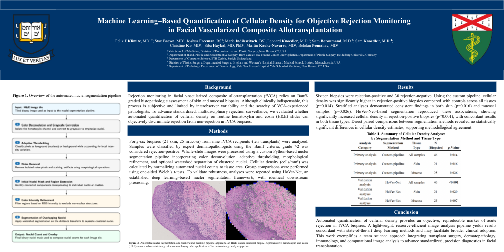
Background: Rejection monitoring in facial vascularized composite allotransplantation (fVCA) relies on Banff-graded histopathologic assessment of skin and mucosal biopsies. Although clinically indispensable, this process is subjective and limited by interobserver variability and the scarcity of VCA-experienced pathologists. To advance standardized, multidisciplinary rejection surveillance, we evaluated whether automated quantification of cellular density on routine hematoxylin and eosin (H&E) slides can objectively discriminate rejection from non-rejection in fVCA biopsies.
Methods: Forty-six biopsies (21 skin, 25 mucosa) from nine fVCA recipients (ten transplants) were analyzed. Samples were classified by expert dermatopathologists using the Banff criteria; grade ≥2 was considered rejection-positive. Whole-slide images were processed using a custom Python-based nuclei segmentation pipeline incorporating color deconvolution, adaptive thresholding, morphological refinement, and optional watershed separation of clustered nuclei. Cellular density (cells/mm²) was calculated by normalizing automated nuclei counts to tissue area. Group comparisons were performed using one-sided Welch’s t-tests. To validate robustness, analyses were repeated using HoVer-Net, an established deep learning–based nuclei segmentation framework, with identical downstream processing.
Results: Sixteen biopsies were rejection-positive and 30 rejection-negative. Using the custom pipeline, cellular density was significantly higher in rejection-positive biopsies compared with controls across all tissues (p=0.014). Stratified analyses demonstrated consistent findings in both skin (p=0.016) and mucosal samples (p=0.026). HoVer-Net–based segmentation reproduced these associations, showing significantly increased cellular density in rejection-positive biopsies (p<0.001), with concordant results in both tissue types. Direct paired comparisons between segmentation methods revealed no statistically significant differences in cellular density estimates, supporting methodological agreement.
Conclusion: Automated quantification of cellular density provides an objective, reproducible marker of acute rejection in fVCA biopsies. A lightweight, resource-efficient image analysis pipeline yields results concordant with state-of-the-art deep learning methods and may facilitate broader clinical adoption. This work exemplifies a team science approach integrating transplant surgery, dermatopathology, immunology, and computational image analysis to advance standardized, precision diagnostics in facial transplantation.